Peyer, Alexander, Girard, Alfred Conrad, 1841-1914, ed. and tr · New York : D. Appleton and company · leaf 370
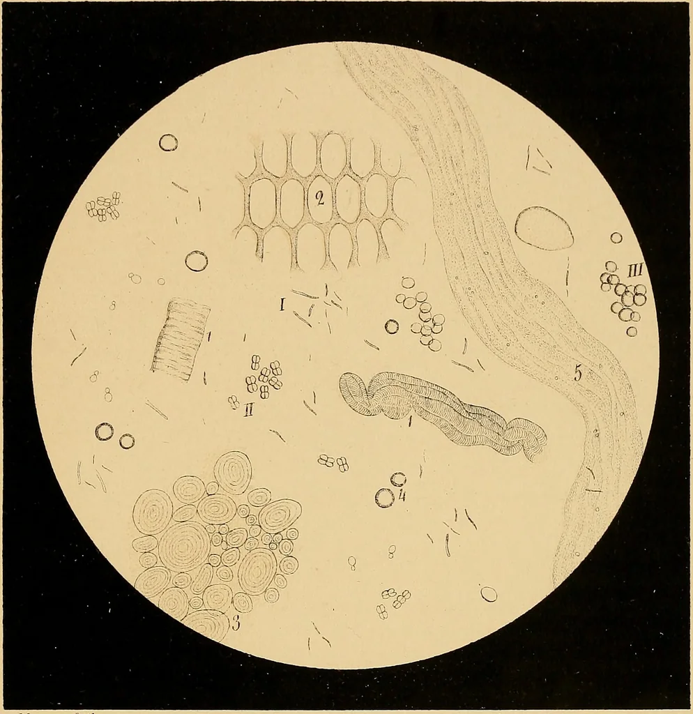
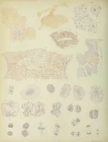
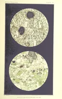
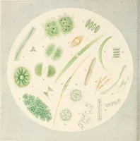
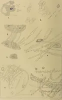
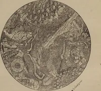
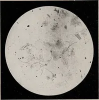
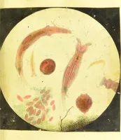
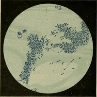
y recognized ifthey have sojourned but a short time in the stomach, but soon thehsemoglobin is disintegrated, and its result, the hsematin, colors thecontents dark brown (coffee-ground vomiting in cancer of stom-ach). The hsemin reaction is then necessary to establish the pres-ence of blood. (See… … Mezger Utk. Vüinii in ciironiG ,q^astric cat-arrh Peyers microscopy. MICROSCOPY OF CONTENTS OF STOMACH. 187 1. Bacteria of the bacillus kind in varioiTS lengths. 2. Sarcina ventricuU. It closely resembles the sarcina nrinse,is only somewhat larger; when it occurs in greater numbers it maybe…
Medical microscopy · Microscopy
Internet Archive · Medical Heritage Library · Public Domain Mark 1.0. Free to use for anything.