Methods in microscopial research; vegetable histology (1905)
Flatters, Abraham · London, Sherratt and Hughes · leaf 146
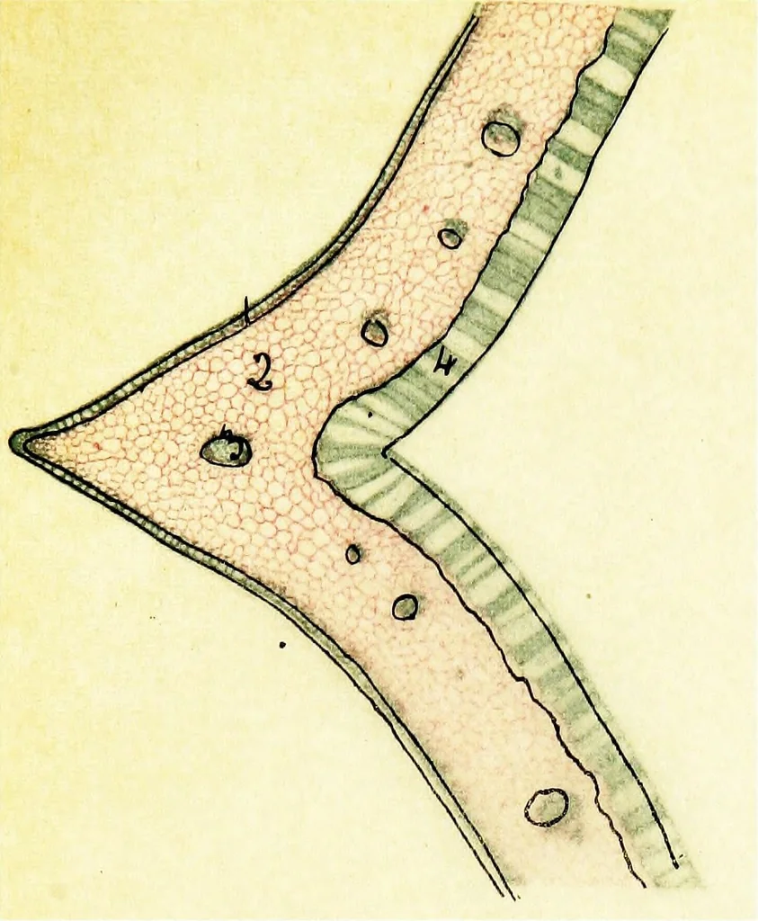
h Chromo-Acetic solution.Cut ^ / 2688 in. Stained with Aniline-picrate and Gossypimine. 1. Palisade layer, continuous on upper and under side. 2. Stomata. 3. Guard cells. 4. Primary ground tissue. 5. Xylem elements of bundle. 6. Phloem elements.7. Small vascular bundles. 8. Oil gland. Fig. 40. Part…
Microscopy · Plant anatomy
Internet Archive · Cornell University Library · Public Domain Mark 1.0. Free to use for anything.