Inside Out Library · Explore
Methods in microscopial research; vegetable histology (1905)
Flatters, Abraham · London, Sherratt and Hughes · leaf 200
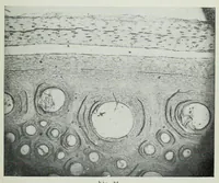
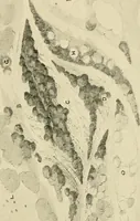
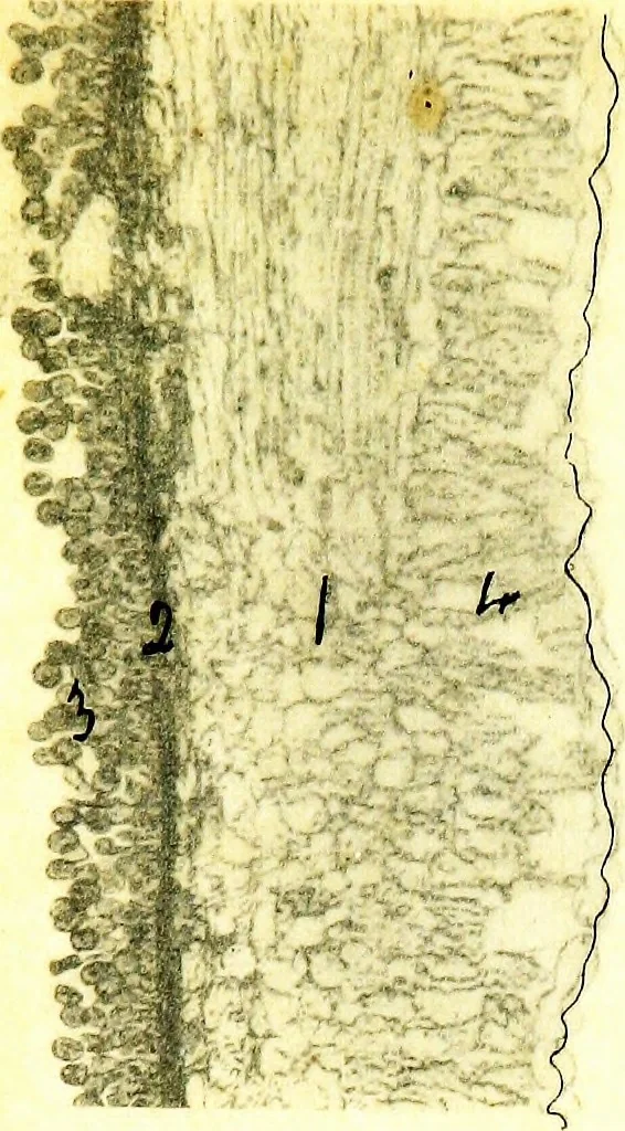 cted with secidiaspores of Puccinia graminis x 36 dia.Fixed with 92% Alcohol. (Unstained.) 1. Ground tissueof leaf. 2. Palisade layer. 3. Mycelium of fungus.4. Fully-developed secidium. 5. Spermogone of unknownfunction. Fig. 73. Vertical section through entire plant of Peziza convexulaX 60 dia.…
Microscopy · Plant anatomy
Internet Archive · Cornell University Library · Public Domain Mark 1.0. Free to use for anything.
More pictures from Methods in microscopial research; vegetable histology
More like this