The microscopy of vegetable foods : with special reference to the detection of adulteration and the diagnosis of mixtures (1906)
Winton, Andrew Lincoln, 1864-1946, Moeller, Josef, 1848-1924 · New York : John Wiley & Sons · leaf 276
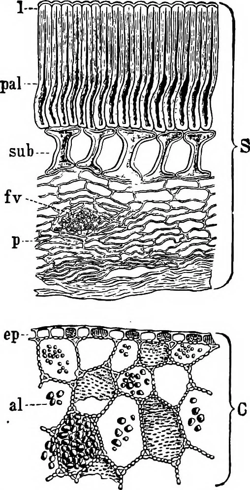
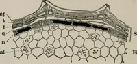
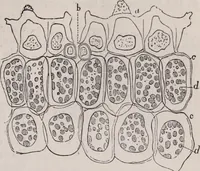
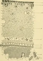
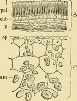
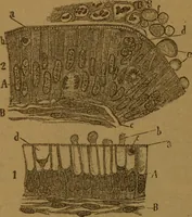
ular chiefly m the mner portion of the cavity. bundle; c cotyledon with ep epi- 2. The Column Cells (Fig. 202, sub; demis and a/aleuronc cells. Xi6o. ^011 (WiNTON.) Fig. 203, t) are 35-70 ,« high, 25-50 ,« broad, hour-glass- or spool-shaped, much constricted in the middle. 3. Parenchyma (Fig. 202,… … 254 LEGUMES. Embryo (C). As noted by Bohmer, the outer epidermal cells ofthe cotyledons are finely porous; these pores, however, are confinedto the radial walls and the edges of the tangential walls. The remainder of the cotyledons consists of Isodiametric cells withmuch swollen, porous walls,…
Plant anatomy · Food adulteration and inspection · Microscopy
Internet Archive · Cornell University Library · Public Domain Mark 1.0. Free to use for anything.