The microscopy of vegetable foods : with special reference to the detection of adulteration and the diagnosis of mixtures (1906)
Winton, Andrew Lincoln, 1864-1946, Moeller, Josef, 1848-1924 · New York : John Wiley & Sons · leaf 306
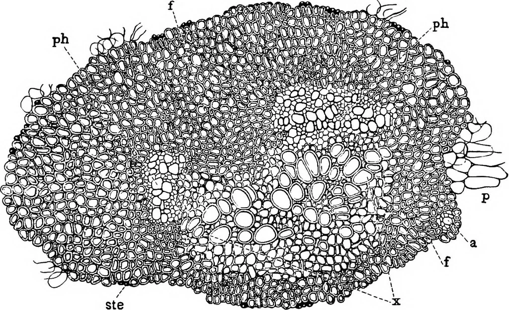
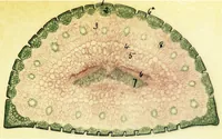
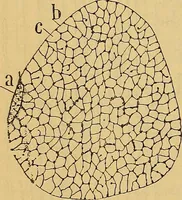
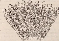
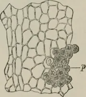
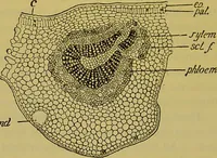
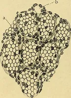
Fig. 230. Cocoanut. In-ner surface of shell withadhering outer spermo-derm. At the left theraphe, from which pro-ceed veins forming a net-work over the surface.X^. (WiNTON.) HISTOLOGY. Pericarp, i. The Epicarp consists of a single layer of rectangularcells with dark contents. … Fig. 231. Cocoanut. Cross section of a large flattened (mesocarp) fiber, ste stegmata;/ sheath of bast fibers; ph two phloem groups; x xylem; p parenchyma of groundtissue; a rudimentary bundle belonging to sriiall branch. X90. (WiNTON.) 2. Mesocarp. The outer portion consists of a ground tissue of…
Plant anatomy · Food adulteration and inspection · Microscopy
Internet Archive · Cornell University Library · Public Domain Mark 1.0. Free to use for anything.