The microscopy of vegetable foods : with special reference to the detection of adulteration and the diagnosis of mixtures (1906)
Winton, Andrew Lincoln, 1864-1946, Moeller, Josef, 1848-1924 · New York : John Wiley & Sons · leaf 431
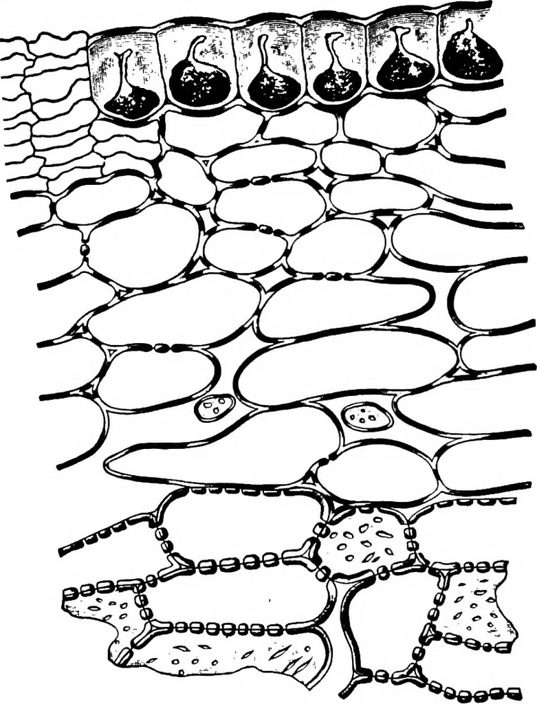
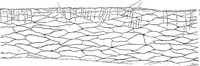
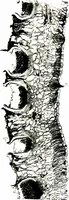
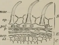
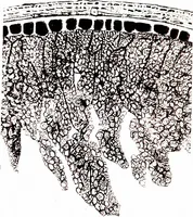
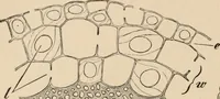
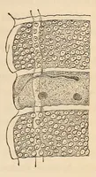
ta andmulticellular hairs. 2. Hypoderm. Moderately thick-walled pitted cells form this layer. 3. Mesocarp. Bundles and latex tubes are scattered through a massof loose parenchyma. 4o8 VEGETABLES. Spermodenn. i. The Palisade Epidermis is strengthened by rodswithout evident branches. 2. Pitied Cells… … Fig. 311. Muskmelon (Cucumis Melo). Cross section of rind. (Moeller.) thickened porous cells intermediate in characters between the corre-sponding cells of the pumpkin and the cucumber. Perispenn, Endosperm, and Embryo are like those of the cucumber. WATERHELON. The watermelon (Citrullus vulgaris…
Plant anatomy · Food adulteration and inspection · Microscopy
Internet Archive · Cornell University Library · Public Domain Mark 1.0. Free to use for anything.