The microscopy of vegetable foods : with special reference to the detection of adulteration and the diagnosis of mixtures (1906)
Winton, Andrew Lincoln, 1864-1946, Moeller, Josef, 1848-1924 · New York : John Wiley & Sons · leaf 507
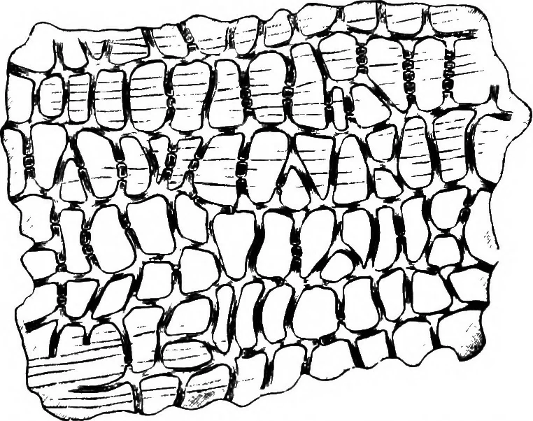
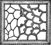
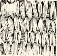
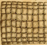
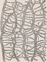
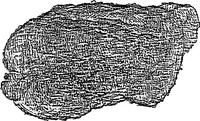
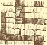
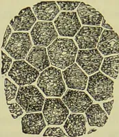
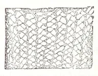
Fig. 417. Mat^ {IlexParaguariensis). Leaf,natural size. (Moel-LER.) 484 ylLKJLOIDAL PRODUCTS.HISTOLOGY. Epidermis (Figs. 418 and 4x9). On both sides the cells are more orless polygonal, with striated cuticle. Along the veins they are arranged … Fig. 418. Mat^. Upper epidermis of leaf, from one of the veins. (Moellee.) side by side in rows. Numerous stomata, larger than the surroundingcells, occur on the lower epidermis. The Mesophyl contains oxalate rosettes. Thick strands of fibersaccompany the bundles.
Plant anatomy · Food adulteration and inspection · Microscopy
Internet Archive · Cornell University Library · Public Domain Mark 1.0. Free to use for anything.