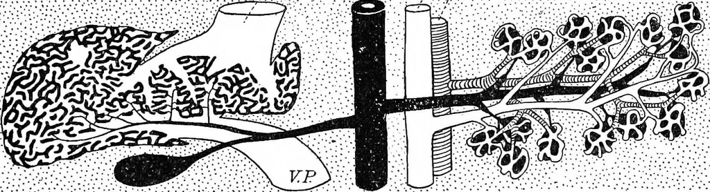
from other vessels and fuse with them. Through theanastomosis of Such sprouts, networks of vessels of small caliber areproduced which have been divided into two types, the sinusoid and capillarytypes. Sinusoids are formed as branches or subdivisions of a single vessel.A vein passing near a… … Fig. 150.—Diagram Showing on the Left the Liver and its Sinusoids ; on the Right the Pancreas and its Capillaries. The connective tissue is represented by dots. Ar., Artery ; Int., intestine ; V., veins ; V. C. I., vena cava inferior; V. P., portal vein. is related in this way to the vitelline…
Histology · Microscopy
Internet Archive · Cornell University Library · Public Domain Mark 1.0. Free to use for anything.