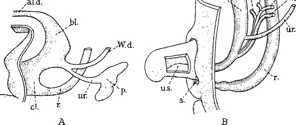
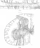
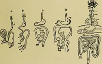
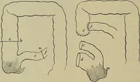
Itarises in two parts, one an outgrowth of the Wolffian duct; and the other,aTmass of dense mesenchyma which is said to be derived from the posteriornephrotomes. In this mesenchyma tubules are formed, which have atone end glomeruh similar to those of the Wolffian body, but smaller. Thetubules… … Fig. 279.—The Development of the Renal Pelvis and Ureter. (After Keibel.)fl, Human embryo of 11.5 mm. (4}^ weeks) ; B, 25 mm, (8J^-9 weeks), a., Anus; al. d., allantoic duct;bl., bladder; cl.,cloaca; M. d., Mullerian duct; r,, rectum ; ur.,ureter; u. s., urogenital sinus ; W. d.,Wolffian duct.…
Histology · Microscopy
Internet Archive · Cornell University Library · Public Domain Mark 1.0. Free to use for anything.