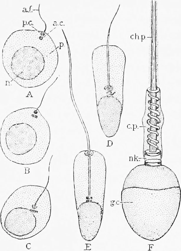
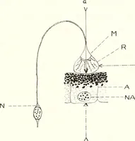
ining rows of granules. Each of them divides intotwo secondary spermatocytes (praespermatids) which are similar cells,though smaller and nearer the lumen. They also have the reducednumber of chromosomes. Every secondary spermatocvte divides intotwo decidedly smaller spermatids, giving them the… … Stages ill iJie trans]on)ialion oj a spermatid into a spermatozoon are shown in the diagram Fig. 307. The twel\-e (?) cliromosomcs of the spermatid disappear in a dense chromatic networlc AAhich becomes an apparently homogeneous mass. Tliis deeply staining nucleus passes to one end of the…
Histology · Microscopy
Internet Archive · Cornell University Library · Public Domain Mark 1.0. Free to use for anything.