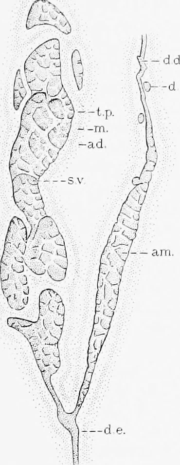
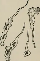
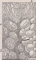
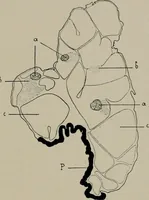
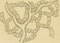
ts.The seminal vesicles grow out from the ductus deferentes at theprostatic ends of their ampullae. Each consists of a number of saccularexpansions arranged along the main outgrowth which is irregularly coiled.The hning of the sacs is honeycombed with folds as shown in Figs. 317and 318. The… … FiG. 317, — Seminal Vesicle anij Ductus Def-erens. (This is natural size.) (After PIberth.) ad., Adventitia; am., atnptiila; d., diverticultini;d. d., ductus duferens; d. e., ductus ejacula-tiiriiis; m., uiusctdan^ ; s. v., sciuinal \ esicle ;t. p.. tunica propria.
Histology · Microscopy
Internet Archive · Cornell University Library · Public Domain Mark 1.0. Free to use for anything.