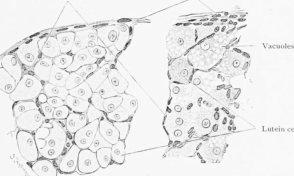
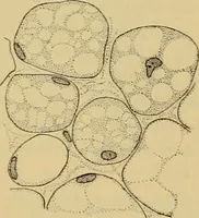
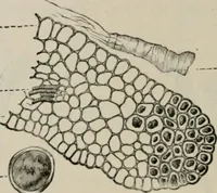
stals, is gradually abscjrbecl. Between thelutein cells there are strands of vascular connective tissue as shown inFig. 334. If pregnancy does not occur the corpus luteum attains itsmaximum development in 12 days and degenerates within a few weeks. Connective tissue septa. Fibrous conneeti\e tissue. … Lutein cells. ^t A B Fig. 334.—A, Portion <.V a CoRPt_-s Luteum of a Rabbit. B, Portion of a Corpus Luteum of a Cat. X 260. In B the lutein cells lia\e become fatty and contain lartce and small vacuoles. Connective tissue increases anrl the lutein cells disintegrate; the newlyformed vessels are…
Histology · Microscopy
Internet Archive · Cornell University Library · Public Domain Mark 1.0. Free to use for anything.