Medical microscopy : a guide to the use of the microscope in medical practice (1898)
Wethered, Frank Joesph, 1860-1928 · Philadelphia : Blakiston · leaf 187
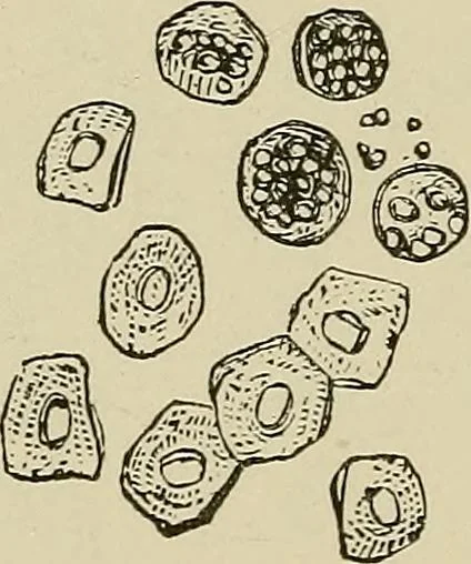
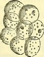
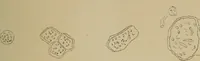
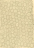
es. Thesingle nucleus is larger, and the protoplasm is more finelygranular. In cases of vesical or renal calculi these cellsare often found in large numbers; they also occur inthe urine of patients suffering from pyelitis. The recognition of renal epithelium (Fig. 29) is of muchmore importance. The… … Renal Epithelium, Healthy and Fatty. The typical cells from the cortical portion consist of acircular nucleus about the size of a red blood corpusclesurrounded by a finely granular protoplasm, the outline ofwhich is generally indistinct. The cells from the straighttubes are flatter and more cubical…
Microscopy · Laboratory Techniques and Procedures · Medical microscopy
Internet Archive · Medical Heritage Library · Public Domain Mark 1.0. Free to use for anything.