The microscope and histology for the use of laboratory students in the anatomical department of Cornell University. by Simon Henry Gage (1891)
Gage, Simon Henry, 1851-1944 · Ithaca, N.Y. : Andrus and Church · leaf 15
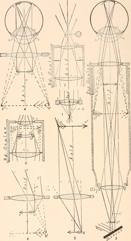
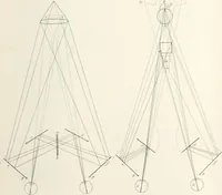
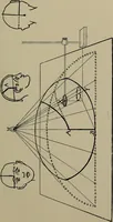
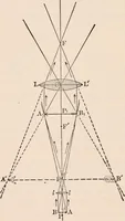
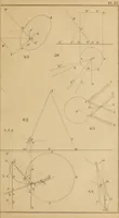
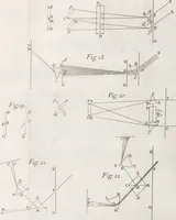
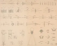
s consists of two optical parts,—an objective, to produce an en-larged, inverted, real image of the object, and an ocular acting in general like asimple microscope to magnify this real image (PI. I, Fig. 5). There is also usuallypresent a mirror, or both a mirror and some form of condenser or… … / 7 / / —L / / \ VL _J3L,M Ri ! oO w l S1 J1 j i ■; i 3: 11 \ - ^^ij^ I EXPLANATION OF PLATE I. In all of the figures, Virtual Images and rays traced backward or produced rays,are indicated by dotted or broken lines, real rays or paths of rays by full or un-broken lines. Fig. i. Sectional view of a…
Microscopy
Internet Archive · Biodiversity Heritage Library · Public Domain Mark 1.0. Free to use for anything.