Carpenter, William Benjamin, 1813-1885 · London : John Churchill · leaf 715
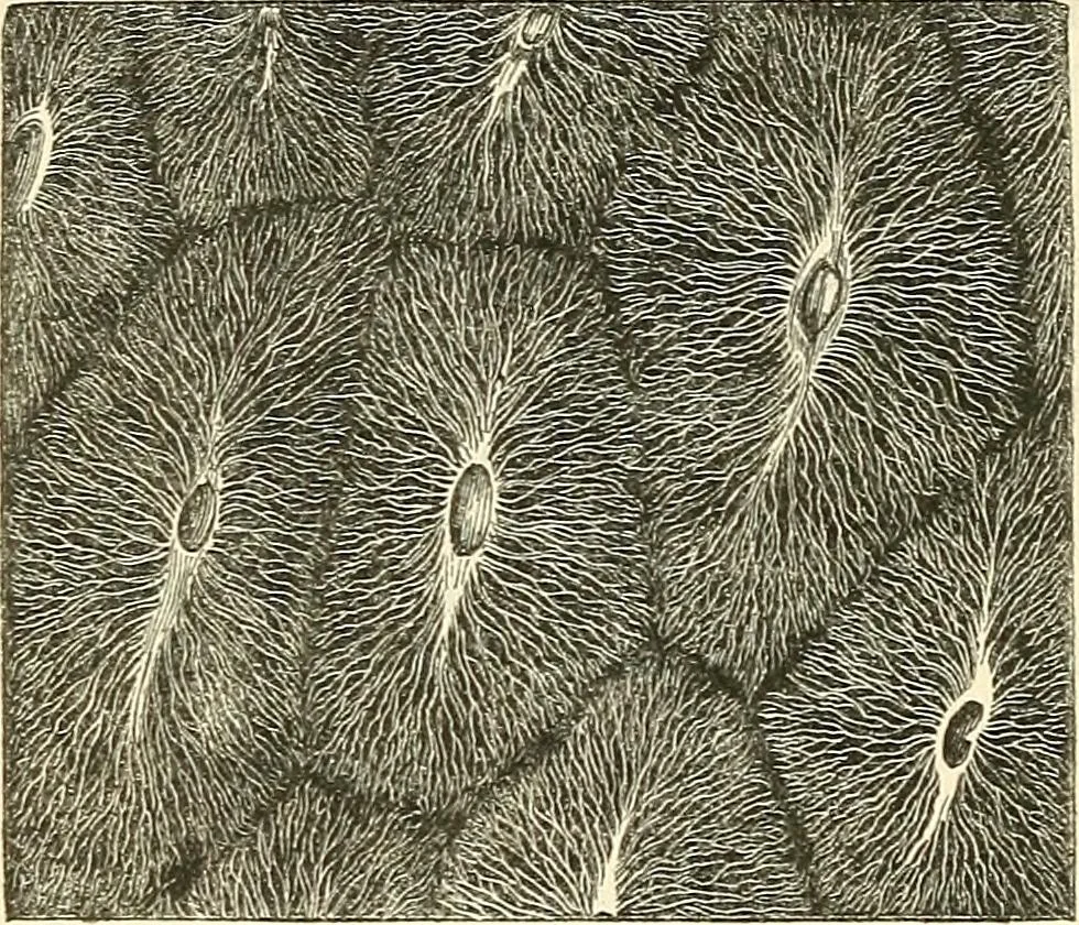
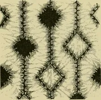
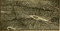
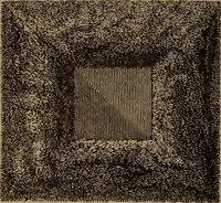
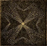
Fig. 302. Perpendicular section of tooth of Lamna, moderatelyenlarged, showing network of medullary canals. Fig. 303. Transverse section of portion of tooth of Prisiis, morehighly magnified, showing orifices of medullary eanals, vnth systemsof radiating and inosculating tubuli. suiTounded by a… … do not enter lacunae, nor is there any concentric annulararrangement around the medullary canals; but each systemof tubuli is continued onwards through its own division of thetooth, the individual tubes sometimes giving-off lateral branches, whilstin other instancestheir trunks bi-furcate. This…
Microscopy
Internet Archive · Medical Heritage Library · Public Domain Mark 1.0. Free to use for anything.