The microscopy of vegetable foods, with special reference to the detection of adulteration and the diagnosis of mixtures (1916)
Winton, Andrew Lincoln, 1864-1946, Moeller, Josef, 1848-1924, Winton, Kate Grace Barber, 1882- · New York, John Wiley & sons, inc.; [etc., etc.] · leaf 291
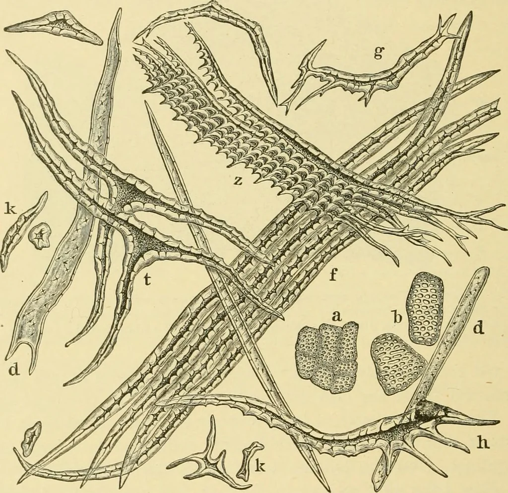
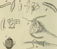
, part of each belonging to the fiber-layer proper, theremainder to a ridge. 272 LEGUMES. In the channels of these outgrowths run the fibro-vascular bundleswith well-marked bast fibers (b), phloem (ph), and xyle.n (xy). 5. Inner Parenchyma (p). Cross sections of partly ripe seeds showa thick inner… … Fig. 224. Peanut. Isolated elements of the pericarp, a anu b cells of the hypoderm;/, z, k, h, t, d and g cells of the fiber layer. X 160. (Winton.) The Spermoderm (Fig. 225, S\ Fig. 226) and perisperm form a thindr) skin which may be readily separated and sectioned either dry in paraf-fine, or wet…
Plant anatomy · Food adulteration and inspection · Microscopy
Internet Archive · Library of Congress · Public Domain Mark 1.0. Free to use for anything.