The microscopy of vegetable foods, with special reference to the detection of adulteration and the diagnosis of mixtures (1916)
Winton, Andrew Lincoln, 1864-1946, Moeller, Josef, 1848-1924, Winton, Kate Grace Barber, 1882- · New York, John Wiley & sons, inc.; [etc., etc.] · leaf 315
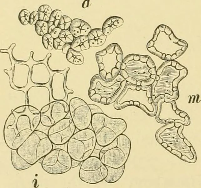
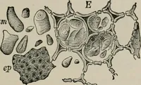
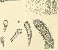
lls with almost colorless walls so stronglythickened that the lumen is scarcely evi-dent. 2. Middle Endocarp (m). The cells in-crease in size and the walls diminish inthickness in the middle layers, the thick-ness of the walls in most of the cells beingmuch less than the breadth of the lumen.Many… … Fig. 242. A alnut {Juglans regia).Tissues of shell, a stone rellsof outer layer; fn stone cellsof middle layer; / parenchymaof inner layer. Xi6o. (Moel- LER.) EUROPEAN IVALNUT. 297 Spermoderm. The seed may be easily sectioned without specialpreparation. The cell structure shoidd be studied after…
Plant anatomy · Food adulteration and inspection · Microscopy
Internet Archive · Library of Congress · Public Domain Mark 1.0. Free to use for anything.