The microscopy of vegetable foods, with special reference to the detection of adulteration and the diagnosis of mixtures (1916)
Winton, Andrew Lincoln, 1864-1946, Moeller, Josef, 1848-1924, Winton, Kate Grace Barber, 1882- · New York, John Wiley & sons, inc.; [etc., etc.] · leaf 625
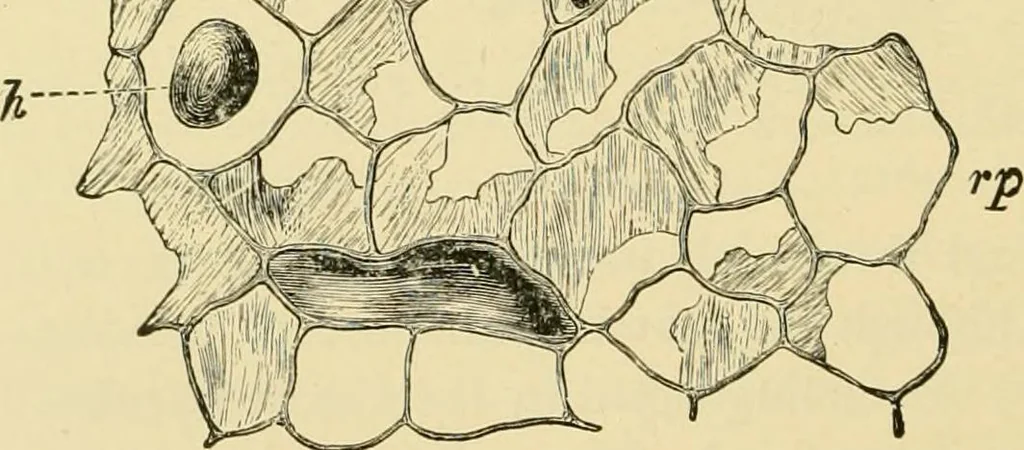
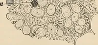
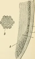
and out, somewhat hard, and have an uneven fracture. Thethickness of the rind exceeds the diameter of the central cyhnder. Crosssections are dotted with dark oil cells and light bundles. HISTOLOGY. 1. The Epidermis (Fig. 520, ep) consists of small polygonal cellsand stomata. T. F. Hanausek notes… … Fig. 520. Galangal {Alpinia officinarum). Outer layers of rhizome in surface view, epepidermis; rp brown parenchyma; g tannin grains; h oil cell. Xi6o. (Moeller.) lumps, but no starch. Further inward the cells have thick porous walls,and contain starch grains. 3. Endodermis (Fig. 521, end). The…
Plant anatomy · Food adulteration and inspection · Microscopy
Internet Archive · Library of Congress · Public Domain Mark 1.0. Free to use for anything.