The microscopy of vegetable foods, with special reference to the detection of adulteration and the diagnosis of mixtures (1916)
Winton, Andrew Lincoln, 1864-1946, Moeller, Josef, 1848-1924, Winton, Kate Grace Barber, 1882- · New York, John Wiley & sons, inc.; [etc., etc.] · leaf 653
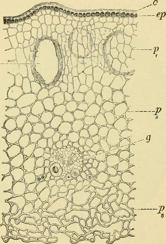
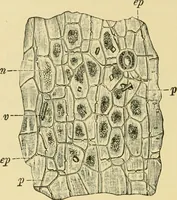
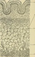
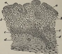
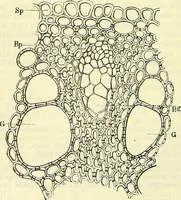
ES AND CONDIMENTS. axis. The structure seen with higher magnification (Fig. 561) is asfollows: 1. Epidermis (ep). The wrinkled epidermis consists of very smallcells with uncommonly thick cuticle (15 /f). In surface view (Fig. 563, A)the cells are sharply polygonal. Stomata occur here and there. 2.… … Fig. 561. Cloves. Cross section through calyx tube. e/> epidermis withe cuticle; />i, ^2,/(gthree forms of parenchyma; o oil cavity; g fibro-vascular bundle with narrow spiralvessel and thick-walled bast fibers. X160. (Moeller.) becoming blue-black with iron chloride are the visible contents.…
Plant anatomy · Food adulteration and inspection · Microscopy
Internet Archive · Library of Congress · Public Domain Mark 1.0. Free to use for anything.