Winton, Andrew Lincoln, 1864-1946, Moeller, Josef, 1848-1924 · New York, J. Wiley & sons; [etc., etc.] · leaf 128
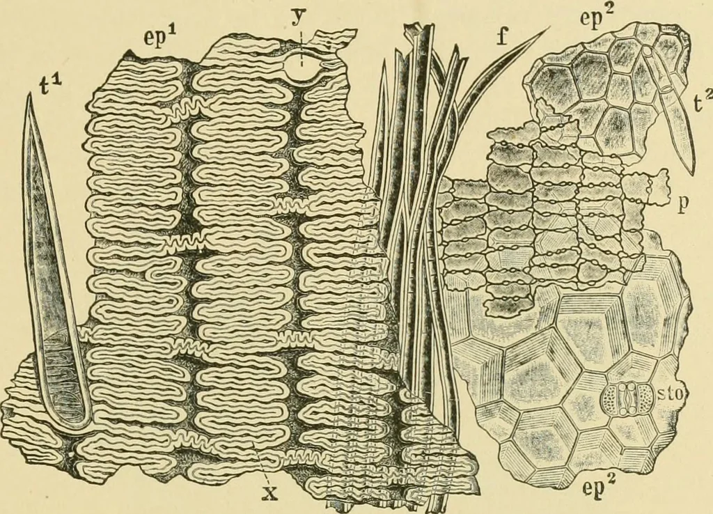
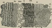
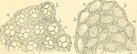
dermis. The inner fibers are thinnerwalled and have outgrowths only on the outer sides. 3. Spongy Parenchyma (p). Two, sometimes more, layers of spongyparenchyma, through which run the bundles, form the mesophyl. Thecells are rectangular, with thin wavy walls. Intercellular spaces occur,not only at… … Fig. 74. Rice. Layers of palet in surface view, ep outer epidermis with x sinuous cells,f hair, and y hair scar; / hypoderm fibers; p spongy parenchyma; ep^ inner epidermiswith 5^0 stoma and i^ hair. X 160. (WiNTON.) show that the cells over the bundles are elongated, but in other parts aremore or…
Food adulteration and inspection · Plant anatomy · Microscopy
Internet Archive · Library of Congress · Public Domain Mark 1.0. Free to use for anything.