Winton, Andrew Lincoln, 1864-1946, Moeller, Josef, 1848-1924 · New York, J. Wiley & sons; [etc., etc.] · leaf 381
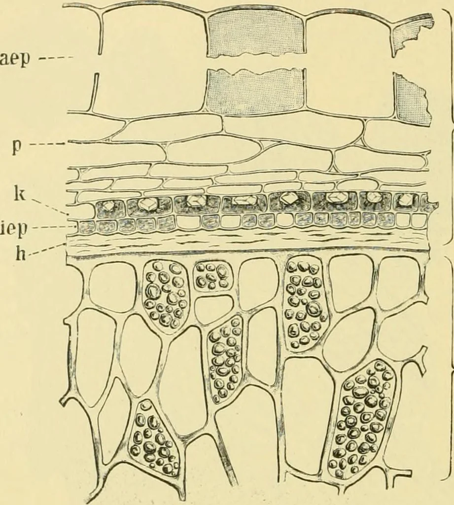
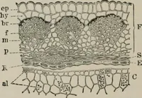
vex. Owingto the great size of the cells, this coat, although but a single cell-layerthick, forms a considerable part of the bulk of the seed. 360 FRUIT. 2. Parenchyma (p). Beneath the mucilage cells are several layersof more or less flattened parenchymatous cells with intercellularspaces* The… … > S E Fig, 273. Red Currant. Seed in cross section. 5 spermoderm consists of aep gelatinousouter epidermis, p parenchyma (nutritive layer), k crystal layer, and iep brown layer(inner epidermis); N perisperm; E endosperm. X300. (Winton.) eters from 8 to 20 «. The middle lamella is colorless, the…
Food adulteration and inspection · Plant anatomy · Microscopy
Internet Archive · Library of Congress · Public Domain Mark 1.0. Free to use for anything.