Winton, Andrew Lincoln, 1864-1946, Moeller, Josef, 1848-1924 · New York, J. Wiley & sons; [etc., etc.] · leaf 42
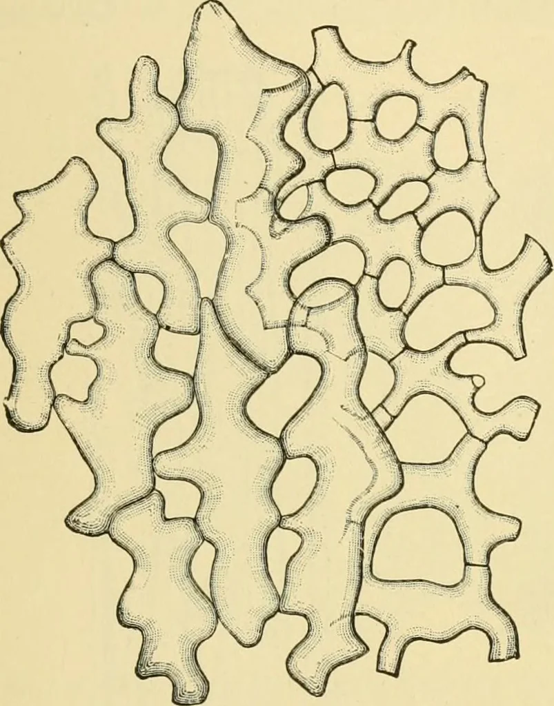
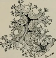
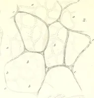
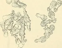
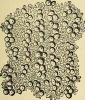
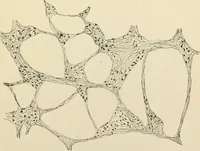
d on the inner surface of thewalls during subsequent growth. Chlorzinc iodine colors the walls yellow THE PRINCIPAL HISTOLOGIC/IL ELEMENTS. 21 or yellow-brown; phloroglucin and hydrochloric acid, pink; aniline sul-phate, deep yellow. Stone Cells (Fig. 4) are isodiametric, or moderately elongated…
Food adulteration and inspection · Plant anatomy · Microscopy
Internet Archive · Library of Congress · Public Domain Mark 1.0. Free to use for anything.