Oral Roentgenology : a Roentgen study of the anatomy and pathology of the oral cavity (1917)
Thoma, Kurt H. (Kurt Hermann), 1883- · Boston, Mass. : Ritter & Company · leaf 108
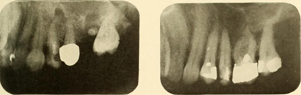
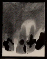
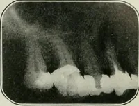
which represents scar-bone that has filled in the place destroyed by the acutesuppuration. Figure 134. Patient: Mr. W. History: He had a very severe attack of acute abscess on the lower first molarmany years ago. Roentgen Examination: Shows root canals partly filled and a radiolucent areaindicating… … Figure 131. Figure 132.