Oral Roentgenology : a Roentgen study of the anatomy and pathology of the oral cavity (1917)
Thoma, Kurt H. (Kurt Hermann), 1883- · Boston, Mass. : Ritter & Company · leaf 184
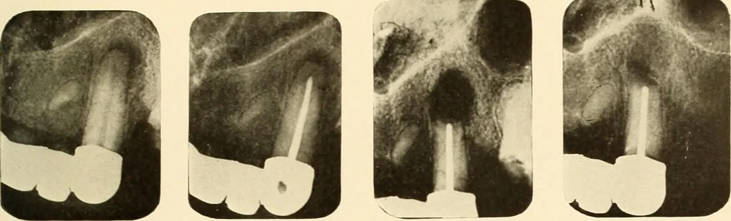
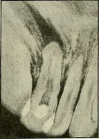
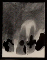
d and filled. Figure 269 shows the condition after apicoectomy was performed. Figure 270 shows the progress of the healing after eight months. Figures 271-274. Figure 271 shows the root canal filling and condition of periapicaltissues before the operation. Figure 272 shows the condition after… … Figure 267. Figure 268. Figure 269. Figure 270. tej