Colwell, Hector Alfred, Russ, Sidney · London, G. Bell and sons · leaf 261
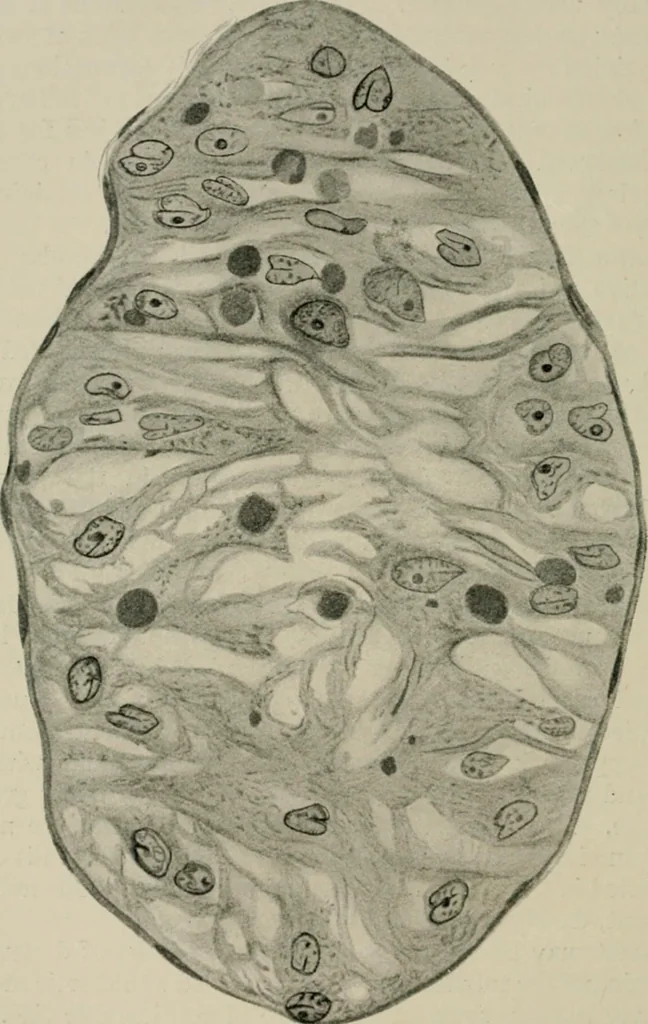
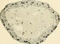
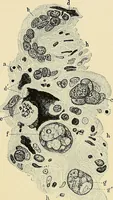
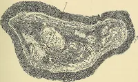
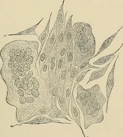
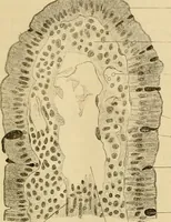
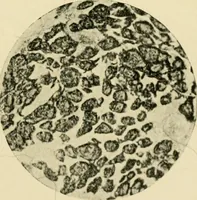
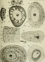
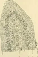
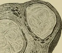
sses, some of which are of considerable size. Theshreddy material which preponderates in the ultimate stage isalso present here, though in smaller amount. Some of the mostcharacteristic changes are illustrated in Figs. 40 and 41. Changes produced in the different elements of the testis. If 242 THE… … Fig. 40.—Seminiferous tubule thirteen days after the application of X rays. All theseminal cells have disappeared. Hxternally the basement membrane is seen. Distributedwithin the tubule are numerous nuclei of cells of Sertoli, the cytoplasm of which foniis acoarse framework, within the meshes of…
X-rays -- Physiological effect
Internet Archive · California Digital Library · Public Domain Mark 1.0. Free to use for anything.