Walsh, David · London : Baillière, Tindall and Cox · leaf 102
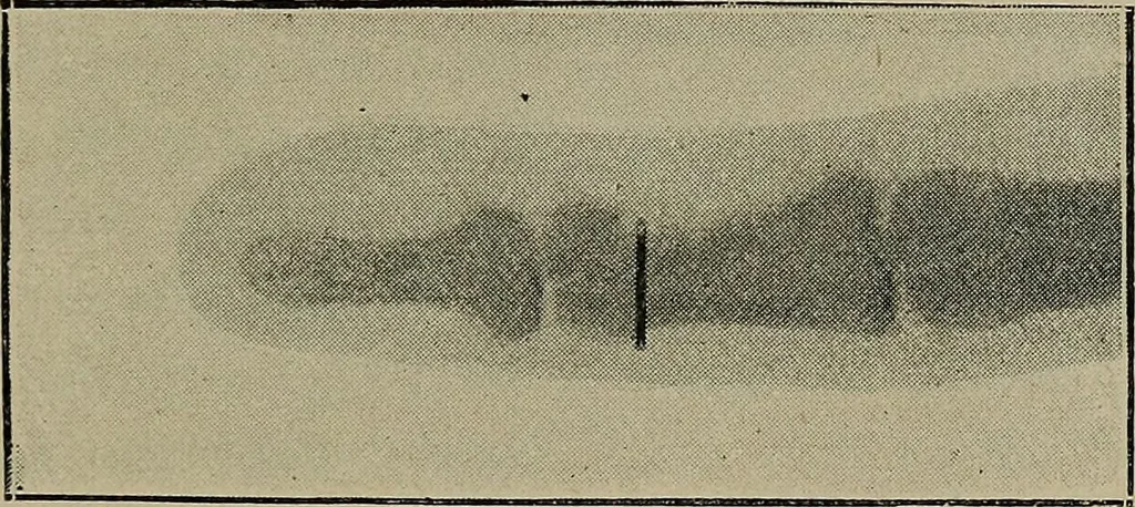
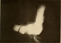
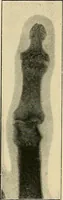
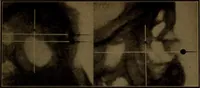
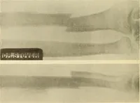
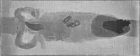
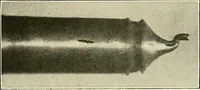
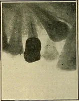
-Sansshowed (1) a slight depression upon the metacarpal bone, doubt-less due to the destruction of the periosteum by the projectile,and (2) the presence in the ball of the thumb of much metallicdebris.. By the courtesy of Dr. Mandras, the photograph is re-produced in Fig. 34. Localization of… … Fig. 35.—Needle in Finger, showing Eye. which shows a needle to be imbedded in the finger. With sosmall an object, the surgeon has no clue as to whether it liesback or front of the digit. A cross-photograph, however, will atonce settle the point. When the fluorescent screen is used, the surgeon…
X-rays · Radiography
Internet Archive · Medical Heritage Library · Public Domain Mark 1.0. Free to use for anything.