Walsh, David · London : Baillière, Tindall and Cox · leaf 136
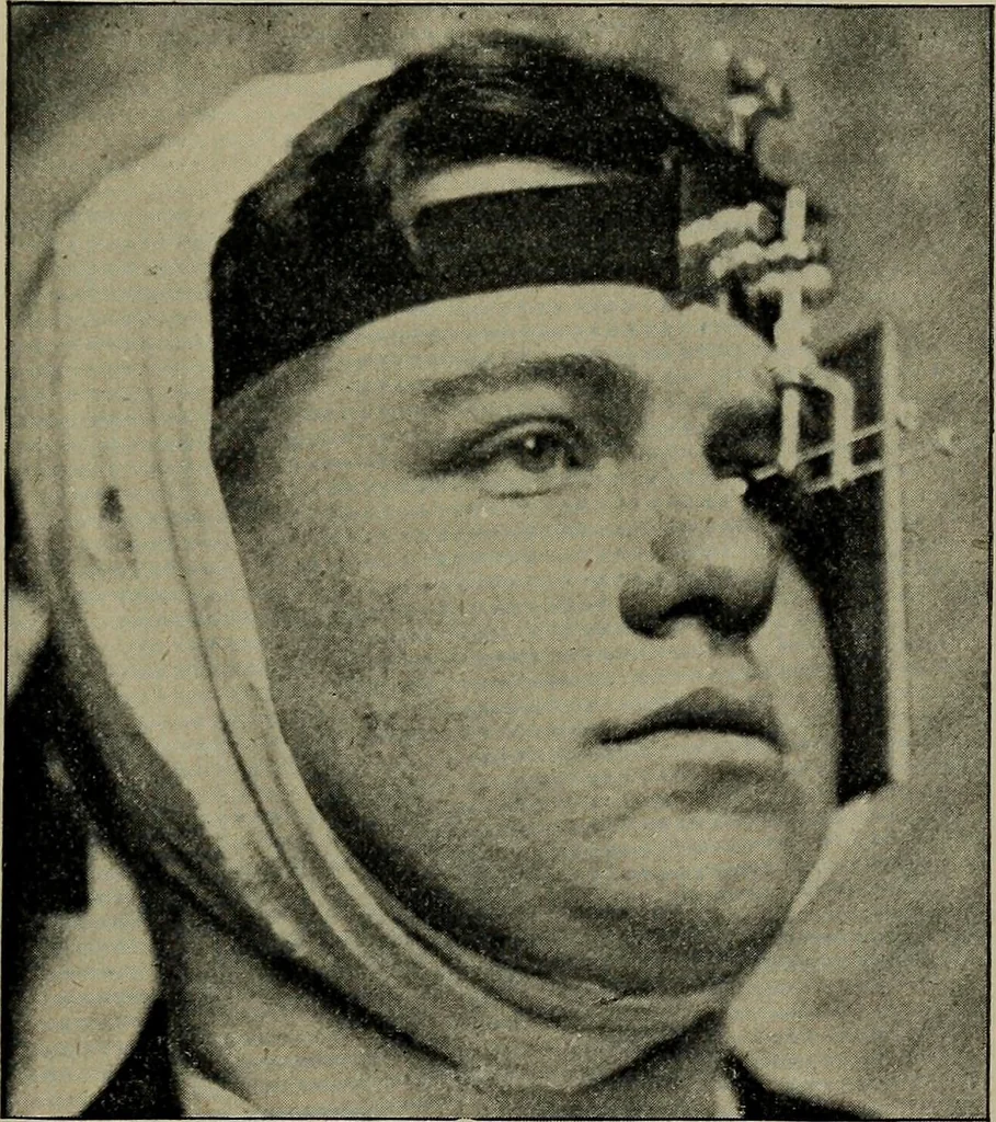
he margin of the malar process of the superior maxillary bone,and the knowledge of the relation of the Crookes tube to thesensitive plate, the location of the foreign body can be closelyestimated. Dr. Stern, of the Philadelphia Polyclinic, however, claims byan original method to have succeeded six… … Fig. 49.—Sweets Localizing Apparatus.From The Practitioner. the fragment in the case of Dr. de Schweinitzs patient, the eyehad actually been cut down upon once, and the magnet appliedthree times with negative results. Good records of small shot and fragments of wire introducedinto orbits were…
X-rays · Radiography
Internet Archive · Medical Heritage Library · Public Domain Mark 1.0. Free to use for anything.