Walsh, David · London : Baillière, Tindall and Cox · leaf 137
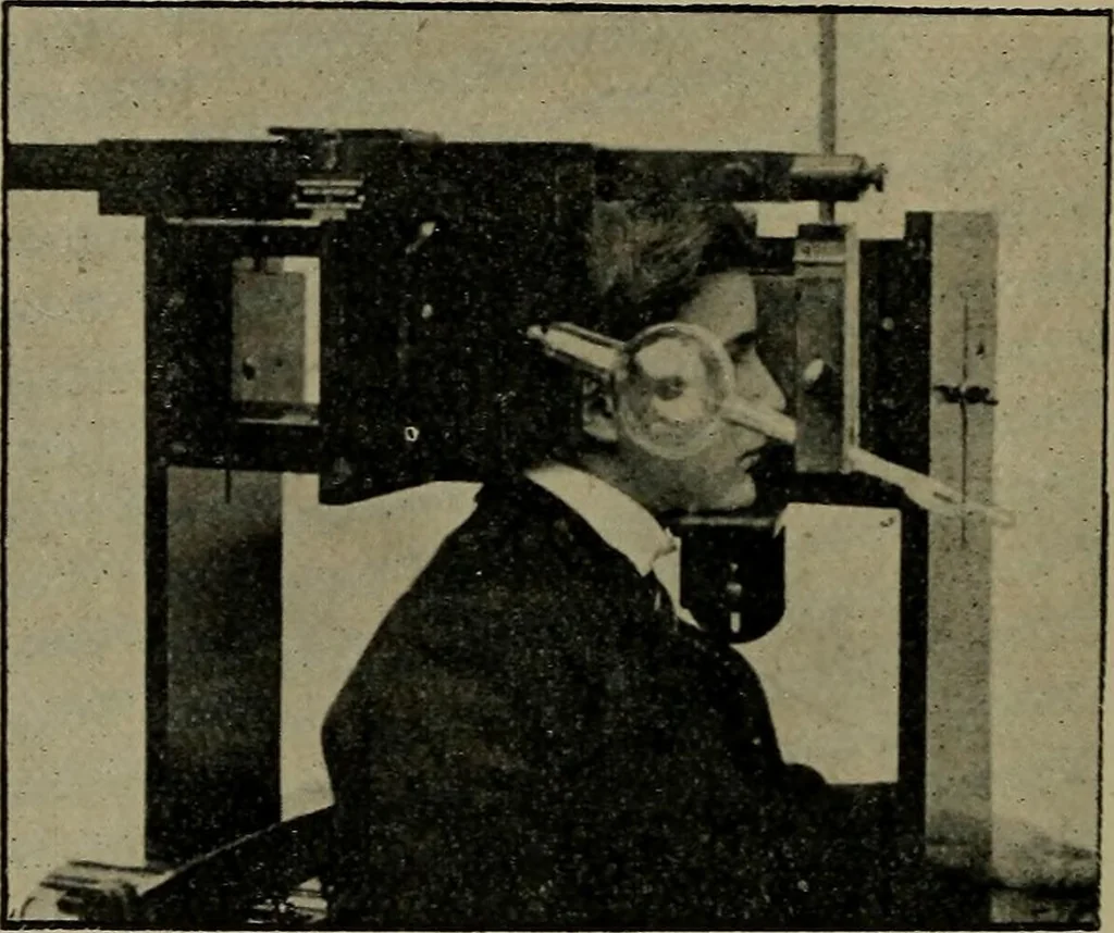
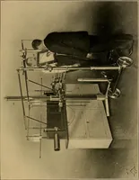
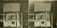
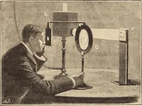
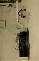
eet* and Dr. Kibbef appear to have led the way;and Dr. Sweet took Eontgen photographs in one or more ofDr. Hansells cases where extraction followed. Dr. Leonardsmethod was published in April, 1898,1 two months later thanthat of Mr. Davidson in England. § An excellent account by Mr. Treacher… … Fig. 50.—Davidsons Eye Appaeatus. carrying two steel rods each, with a rounded ball at the end.These balls are placed at a known distance from the eyeball, onepointing to the centre of the cornea, the other to the externalcanthus, both parallel to the visual line and perpendicular to theplate. 1…
X-rays · Radiography
Internet Archive · Medical Heritage Library · Public Domain Mark 1.0. Free to use for anything.