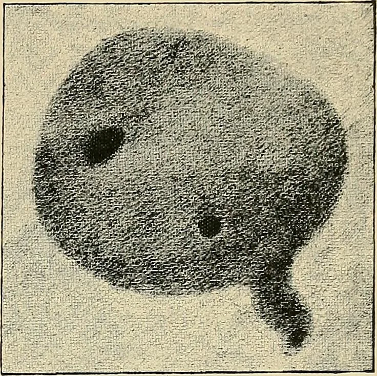
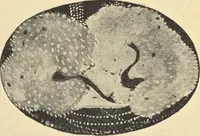
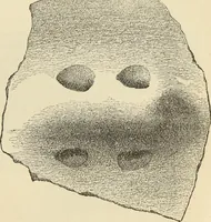
living human eye he used small semicircular sensitizedplates, as that shape allowed the largest possible area to be insertedat the inner angle of the eye opposite the lachrymal bone. Byrolling the eye inwards or outwards, a large part of the globecould be brought within range of the rays. Rotation,… … Fig. 82.—Pellet and Fragment of Lead in Human Eye. Van Duyse. minal placed so as to point exactly to the antero-posterior axisof the centre of the cornea. In this way he obtained localizingradiograms of a foreign body(a sequin) placed on the con-junctiva of his own eyeball. In February, 1897, two…
X-rays · Radiography
Internet Archive · Medical Heritage Library · Public Domain Mark 1.0. Free to use for anything.