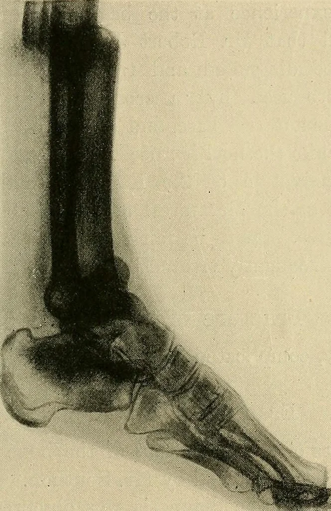
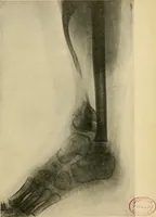
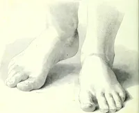
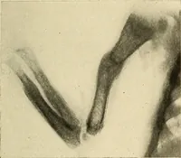
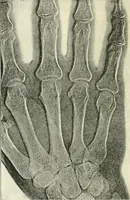
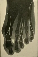
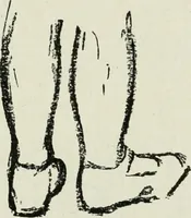
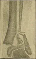
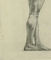
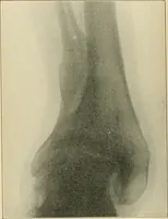
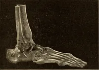
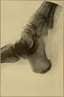
ig. 103.—Fracture of Tibia and Fibula ; Three Months after Injury jno External Deformity. Dr. Lows case. MEDICAL AND SURGICAL APPLICATIONS 223 A helpful classification of fractures about the ankle-joint is thatof Stimson, modified by Barnard by the addition of Dupuytrensfracture, as follows : 1.… … Fig. 104. — Bones of Foot showing Atkophy and Pencilled Outlinefollowing fracture of tlbia and flbtjla. Mr. Robert Joness case. Radiographed by Dr. D. Morgan. Potts fracture is, of course, by far the commonest of theseinjuries, while Dupuytrens occurs only once in about a hundredcases of fracture…
X-rays · Radiography
Internet Archive · Medical Heritage Library · Public Domain Mark 1.0. Free to use for anything.