Sthohr's histology : arranged upon an embroyological basis (1906)
Sthohr, Philipp, 1849-1911, Lewis, Frederic Thomas, 1875 · Philadelphia : P. Blakiston's Son & Co. · leaf 390
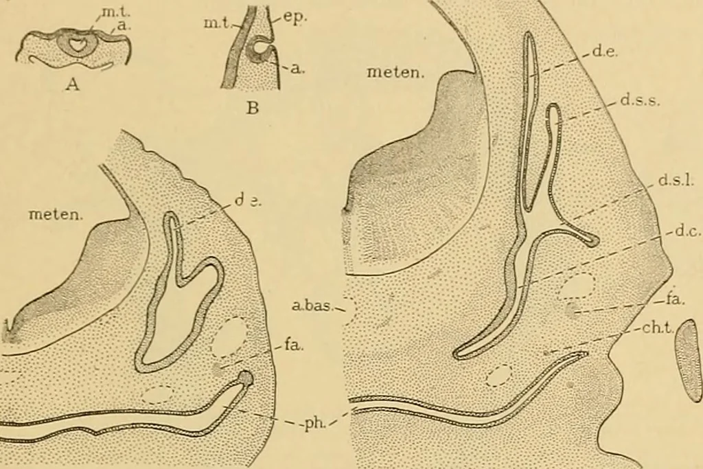
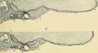
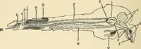
on becomes enlarged to form the endolymphaticsac. The duct is seen in section in Fig. 429, C, and its upper end projectsabove the rest of the vesicle in Fig. 430, A. In the adult it terminatesjust beneath the dura. In two places the medial and the lateral walls of the upper half of thevesicle… … Fig. 429.—Sections of Rabbit Embryos to show the Development of the Ear. X9. A, () days. 3.8 mm.: B, 10 days, 3.4 mm.; C, 12^ days, 7.5 mm.; D, 14 days, 10 mm. a.. Ectodermalepithefium which forms the membranous internal ear; a. bas., basilar artery; ch. t., chorda tympani;d. c, cochlear duct; d.…
Histology · Microscopy
Internet Archive · Public Domain Mark 1.0. Free to use for anything.