The microscopy of vegetable foods, with special reference to the detection of adulteration and the diagnosis of mixtures (1916)
Winton, Andrew Lincoln, 1864-1946, Moeller, Josef, 1848-1924, Winton, Kate Grace Barber, Mrs · New York, John Wiley & sons, inc.; [etc., etc.] · leaf 103
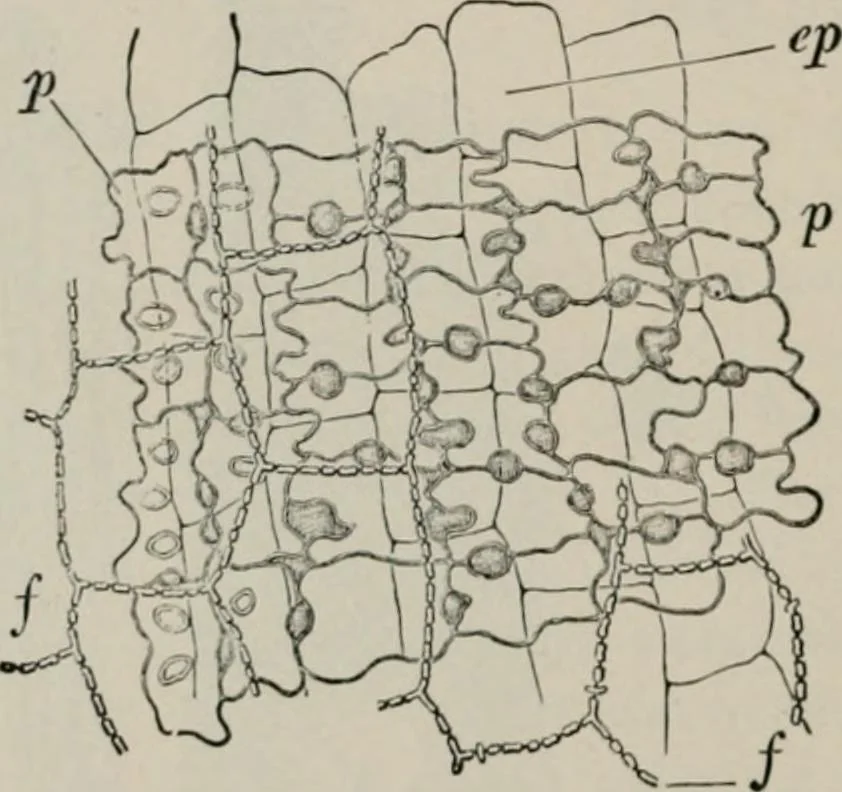
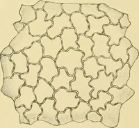
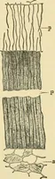
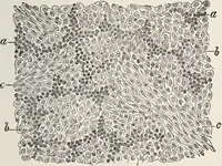
, convoluteddouble walls of the long cells appear to be of uniform structure; but ontreatment with alkali, the zigzag middle lamella separating adjoiningcells is clearly evident. 2. Hypoderm (Fig. 44; Fig. 45, /). One to three layers of fibers withthick, porous walls, underhc the epidermis. 3.… … Fig. 46. Barley. Surface view of p spongy paren-chyma of palet, ep inner epidermis of palet,and/cpicarp. X300. (Moellek.)