The microscopy of vegetable foods, with special reference to the detection of adulteration and the diagnosis of mixtures (1916)
Winton, Andrew Lincoln, 1864-1946, Moeller, Josef, 1848-1924, Winton, Kate Grace Barber, Mrs · New York, John Wiley & sons, inc.; [etc., etc.] · leaf 138
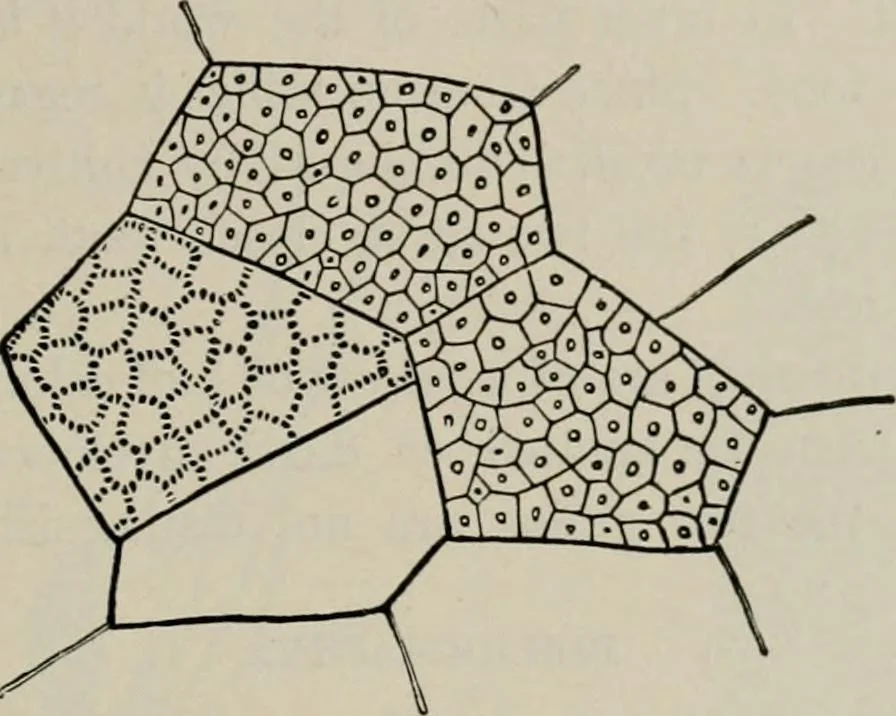
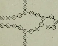
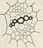
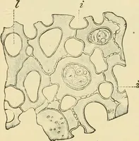
ight end walls. Both forms havesmooth outer walls and arc without loiorid contents. COMMON MILLET. Il7 The Hypoderm Fibers, rectangular parenchyma cells without inter-cellular spaces, and the inner epidermis also of rectangular cells, arethe same as in the glumes and palet of Setaria. The Caryopzis… … Fig. 84, Common Millet {Panicnm mUmceum). Starch cells of endosperm showing (atthe left) beaded network remaining after treatment with alkaU. (VOGL.) with alkali the starch grains dissolve, leaving a beaded network corre-sponding to the form of the grains (Fig. 84). DIAGNOSIS. The chief products of…