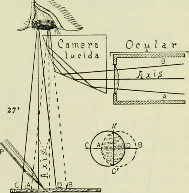
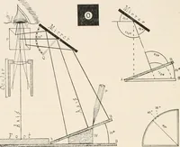
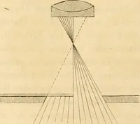
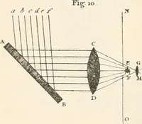
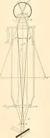
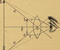
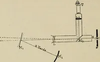
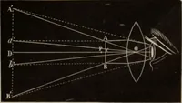
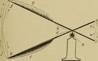
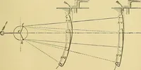
he pupil of the eye. Axis, Axis. Axial rays Jrom the•microscope and from the drawing sur-face (Ch. V). Camera Lucida. A section of thequadrangular prism showing thecourse of the rays in the p> ism fromthe microscope to the eye. As the raysare twice reflected, they have the samerelation on… … Fig. 92 A D, A D. Overlapping portion of the two fields, where both the microscopicimage and the drawing surface, pencil, etc., may be seen. It is represented by theshaded part in the overlapping circles at the right. Ocular. The ocular of the microscope. P. The draiving pencil. Its point is shown…
Microscopy
Internet Archive · Medical Heritage Library · Public Domain Mark 1.0. Free to use for anything.