Methods in microscopial research; vegetable histology (1905)
Flatters, Abraham · London, Sherratt and Hughes · leaf 116
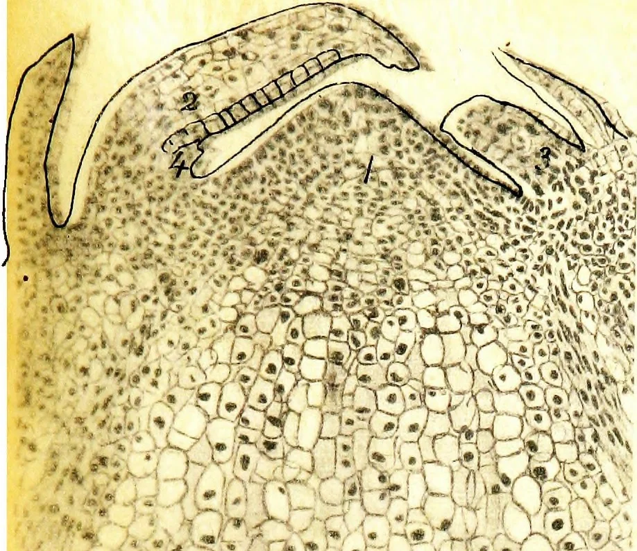
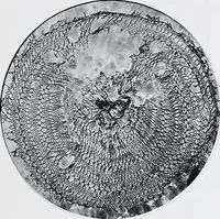
se section, two inches below growing point. Stemof Pine X 35 dia. Tissue fixed aaid stained as in Fig. 20.1. Cortical tissue. 2, Phloem elements. 3. Cambiformtissue. 4. Xylem elements, after the commencement ofsecondary thickening. 5. Ilesin passage. 6. Central groundtissue. 7. Developing branch.…
Microscopy · Plant anatomy
Internet Archive · Cornell University Library · Public Domain Mark 1.0. Free to use for anything.