Inside Out Library · Explore
Methods in microscopial research; vegetable histology (1905)
Flatters, Abraham · London, Sherratt and Hughes · leaf 128
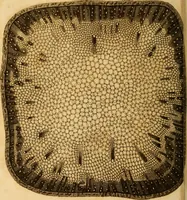
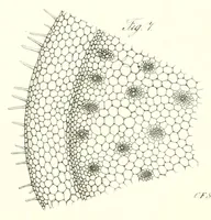
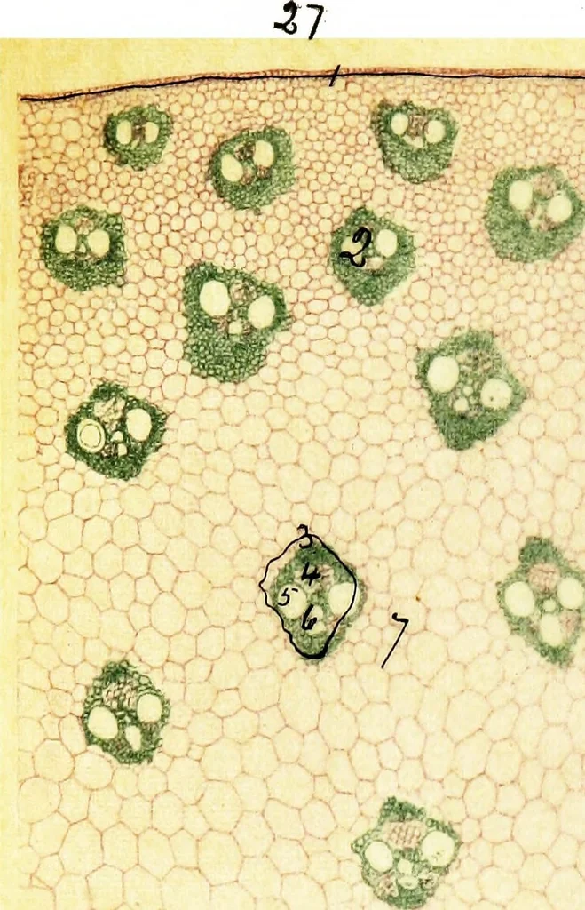 itted vessels. 3. Annular vessel. 4. Spiral vessel. Fig. 29. Transverse section through Rachis of Fern. Pterisaquilina x 40 dia. Fixed in Chromo-Acetic solution.Staining as in above figures. 1. Primary ground tissue.2. Endodermis. 3. Bast sheath. 4. Bast. 5. Scalariformvessels. 6. Spiral vessels.… … ^f
Microscopy · Plant anatomy
Internet Archive · Cornell University Library · Public Domain Mark 1.0. Free to use for anything.
More pictures from Methods in microscopial research; vegetable histology
More like this