Inside Out Library · Explore
Methods in microscopial research; vegetable histology (1905)
Flatters, Abraham · London, Sherratt and Hughes · leaf 86
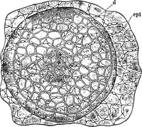
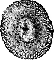
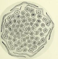
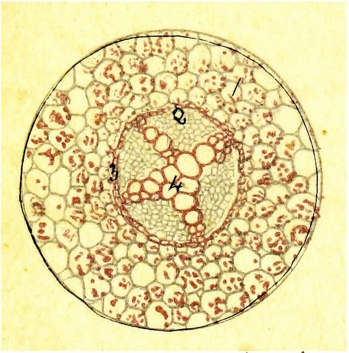 Stained Hsematoxylinand Safranin. 1. Primary ground tissue. 2. Endodermis. 3. Large pitted vessels. 4. Phloem elements, stained purple. 5. Xylem elements, stained red. 6. Developing lateralrootlets. 7. Central axis of root. Fig. 4. Transverse section. Root of Zea mais x 35 dia. Fixedin 92% Alcohol.… … J
Microscopy · Plant anatomy
Internet Archive · Cornell University Library · Public Domain Mark 1.0. Free to use for anything.
More pictures from Methods in microscopial research; vegetable histology
More like this