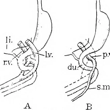
lt. In the embryo these veins are prolonged posteriorly(in part) as the right and left subcardinal veins, shown in the cross section.Fig. 248. This figure indicates also that the liver fuses with the dorsalwall of the abdominal cavity on the right side of the body (at x). No bor-responding adhesion… … Fig. 247.The formation of the portal veinp. v., from the right and leftvitelline veins, r. V. and I. v. ;du., duodenum; li,, liver; s.m. v.,superior mesenteric vein. Digitized by Microsoft® LIVER. 221 the azygos vein, now passes to the viteUine portion of the inferior vena cava.The original…
Histology · Microscopy
Internet Archive · Cornell University Library · Public Domain Mark 1.0. Free to use for anything.