Dental and oral radiography : a text book for students and practitioners of dentistry (1916)
McCoy, James David · St. Louis : Mosby · leaf 145
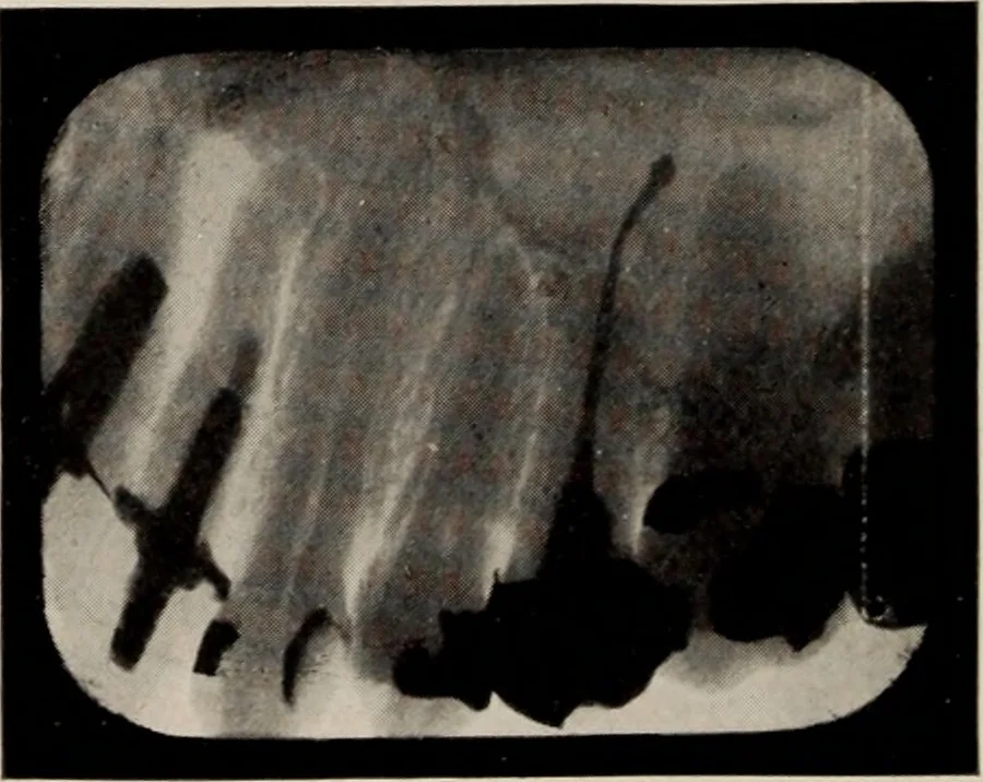
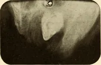
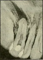
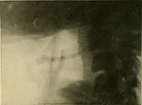
Fig. 68. Small wires introduced into root canals to determine their length orthe extent to which they have been opened, are easily differentiated fromroot canal fillings or tooth structure. … Fig. 69. Root canal filling material forced beyond the root apex of an uppersecond bicuspid. tions appear npon the plate as dark areas owingto the fact that the rays pass through them moreeasily, and effect the emulsion of the plate to a INTERPRETATION OF RADIOGRAPHS 143
Radiography, Dental · X-rays · Teeth · Mouth
Internet Archive · Public Domain Mark 1.0. Free to use for anything.