Medical microscopy : a guide to the use of the microscope in medical practice (1898)
Wethered, Frank Joesph, 1860-1928 · Philadelphia : Blakiston · leaf 171
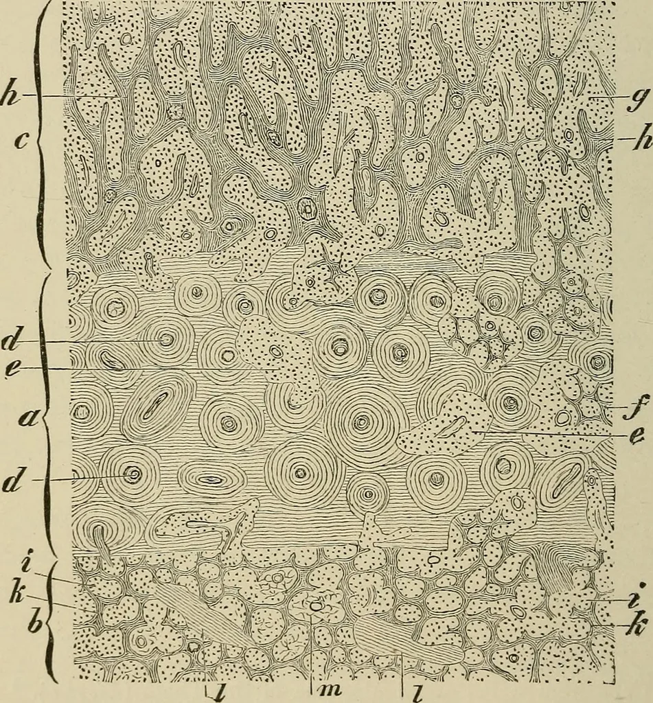
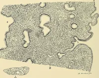
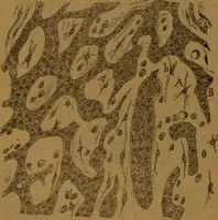
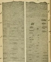
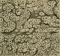
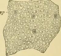
opically, the commoner forms have practicallythe same structure as ordinary hyaline cartilage. In many * Figures 20, 21, 22, 23, 25, 26, from Zieglers Pathological Ana-tomy, are inserted with the kind permission of Messrs. Macmillan&Co. i56 MEDICAL MICROSCOPY. the cartilage cells are more… … Section from an Osteoid Chondroma of the Humerus (Ziegler). cortical layer ; b, medullary spaces or cancelli; c, periosteal growth ; d, normalHaversian canals; e, Haversian canals distended with cartilage, which atf contains a core of new bone ; g, cartilage developed from periosteum whichat h…
Microscopy · Laboratory Techniques and Procedures · Medical microscopy
Internet Archive · Medical Heritage Library · Public Domain Mark 1.0. Free to use for anything.