The microscope and histology for the use of laboratory students in the anatomical department of Cornell University. by Simon Henry Gage (1891)
Gage, Simon Henry, 1851-1944 · Ithaca, N.Y. : Andrus and Church · leaf 19
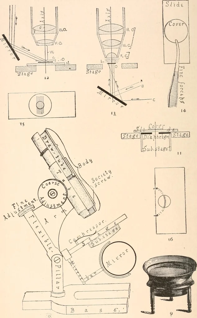
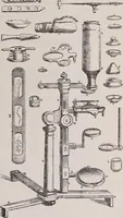
m false color. The field-lensalso aids in rendering the field flat. EL. Eye-lens. FL. Field-lens. Fig. 7. Sectional view of a Huygenian ocular {Hg. ocular), to show the forma-tion of the Eye-Point. Axis. Optic axis of the ocular. D. Diaphragm of the ocular. E L. Eye-lens.F L. Field-lens. E P.… … EXPLANATION OF PLATE II. Fig. 9. Tripod Magnifier. The frame holding the lenses may be raised or low-ered (focused) by screwing it up or down in the outside ring. Fig. 10. Stand. That is the mechaical parts of a simple form of compound mi-croscope, with the names of the parts written upon them.…
Microscopy
Internet Archive · Biodiversity Heritage Library · Public Domain Mark 1.0. Free to use for anything.
![An illustration from Eighty Years' Progress of the United States: a family record of American industry, energy and enterprise ... By eminent literary men [C. L. Flint and others] ... Embellished with ... engravings, etc (1868)](https://img.insideoutlibrary.com/img/200/bl-bl_003734531_1-452-1.webp)