Carpenter, William Benjamin, 1813-1885 · London : John Churchill · leaf 403
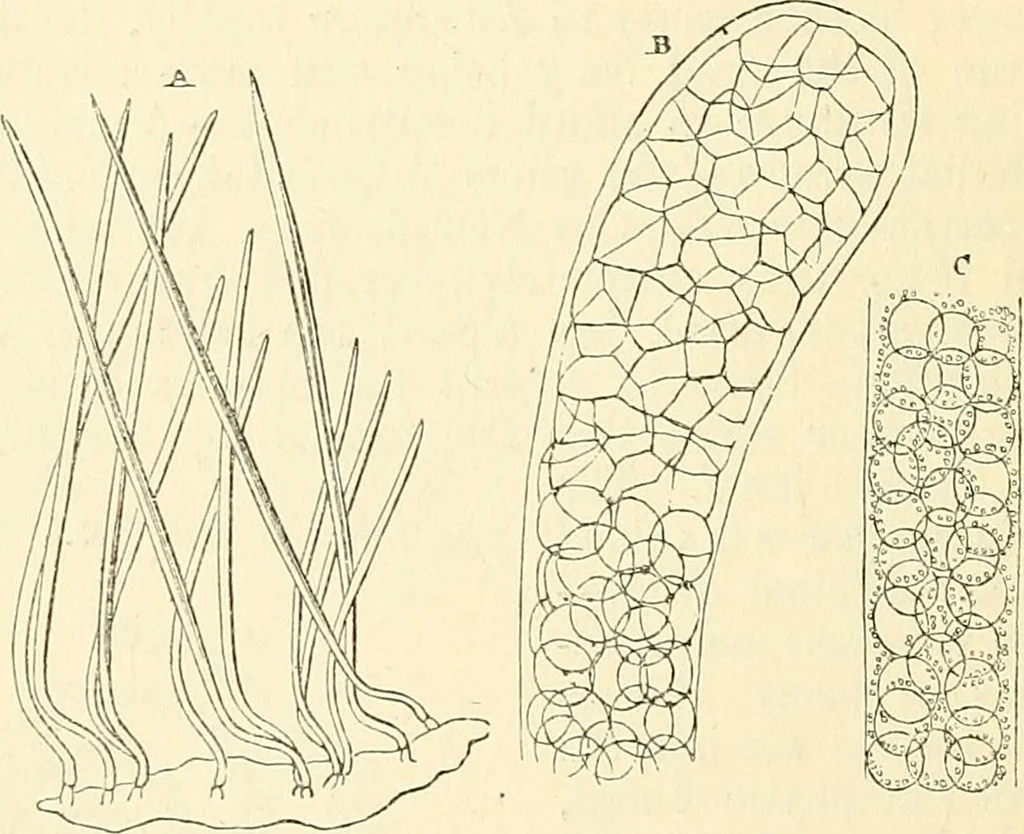
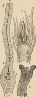
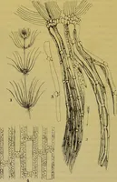
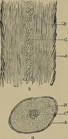
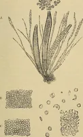
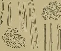
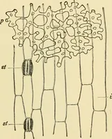
secondary cell. 380 MICROSCOPIC STUUCTURE OP HIGHEE CEYPTOGAMIA. consists of a single long tubular cell, wMch. sometimes growsin a spiral mode (Kg. 121), sometimes straight and tapering(Eig. ].22, a); in its young state, the cell contains a transparentprotoplasma, with granules and globules of… … structure of Enterobr^tis:—A, growth of U. attenuatus, frommucous membrane of stomach of Fassidus; b, dilated extremity ofprimary cell ofE. elegans, tilled with secondary cells, which, near itstermination, become mutually flattened by pressure; c, lower por-tion of the same filament, contaming-…
Microscopy
Internet Archive · Medical Heritage Library · Public Domain Mark 1.0. Free to use for anything.