The microscopy of vegetable foods, with special reference to the detection of adulteration and the diagnosis of mixtures (1916)
Winton, Andrew Lincoln, 1864-1946, Moeller, Josef, 1848-1924, Winton, Kate Grace Barber, 1882- · New York, John Wiley & sons, inc.; [etc., etc.] · leaf 169
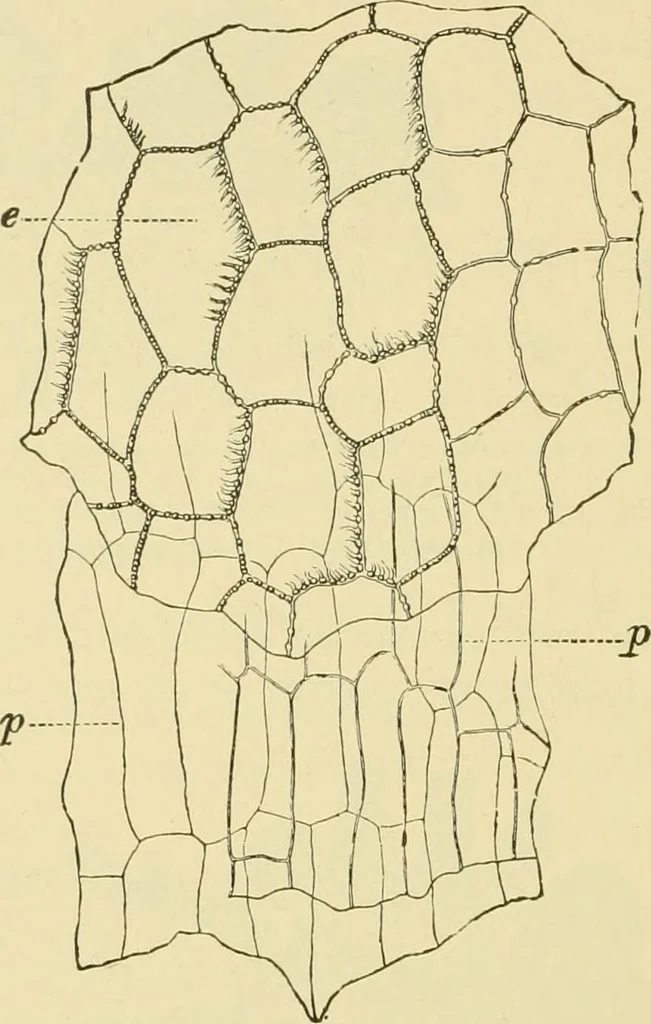
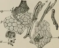
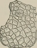
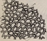
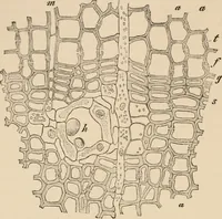
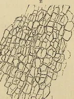
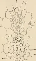
e view. Xi6o. (Moeller.) ous fine warts. They contain a brown substance which is not removedby dilute alkah even on boihng. 2. Parenchyma (Figs. 118 and 120, p). Beneath the epidermis areone or more layers of parenchyma cells with somewhat thickened, brown ISO IVEED SEEDS. walls. These, like the… … Fig. 120. Cockle. Inner layers of spermoderm in surface view, p parenchyma; e innerepidermis. X160. (Moeller.) describe this layer as the inner epidermis of the spermoderm; Vogl,however, regards it as perisperm. Endosperm (Fig. 118, E). The large cells contain highly character-istic, oval-fusiform,…
Plant anatomy · Food adulteration and inspection · Microscopy
Internet Archive · Library of Congress · Public Domain Mark 1.0. Free to use for anything.