The microscopy of vegetable foods, with special reference to the detection of adulteration and the diagnosis of mixtures (1916)
Winton, Andrew Lincoln, 1864-1946, Moeller, Josef, 1848-1924, Winton, Kate Grace Barber, 1882- · New York, John Wiley & sons, inc.; [etc., etc.] · leaf 263
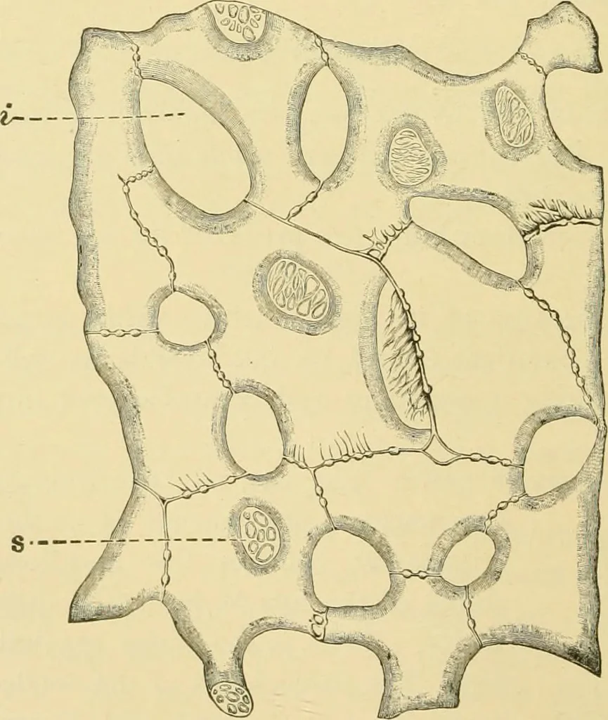
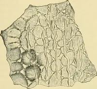
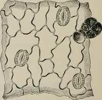
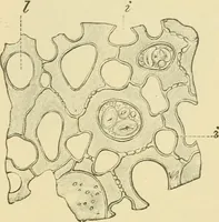
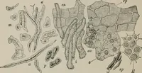
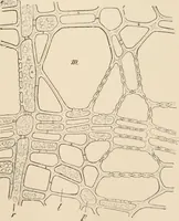
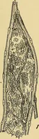
Fig. 191. Pea. Elements of spermoderm in surface \-iew. p palisade cells; t hour-glasscells (subepidermal layer); e parenchyma. X160. (Moeller.) common coffee adulterant in the United States consists of pea hullsmade into pellets with molasses and other ingredients. I … Fig. 192. Pea, Outer layers of spongy parenchyma, i intercellular space; s porousmembrane at end of arm. (Moeller.) The elements of the hull may be studied in section (Fig. 189), orin surface view (Fig. 191) after scraping with a scalpel. Isolation of PEA. LENTIL. 245 the palisade and column cells…
Plant anatomy · Food adulteration and inspection · Microscopy
Internet Archive · Library of Congress · Public Domain Mark 1.0. Free to use for anything.