The microscopy of vegetable foods, with special reference to the detection of adulteration and the diagnosis of mixtures (1916)
Winton, Andrew Lincoln, 1864-1946, Moeller, Josef, 1848-1924, Winton, Kate Grace Barber, Mrs · New York, John Wiley & sons, inc.; [etc., etc.] · leaf 251
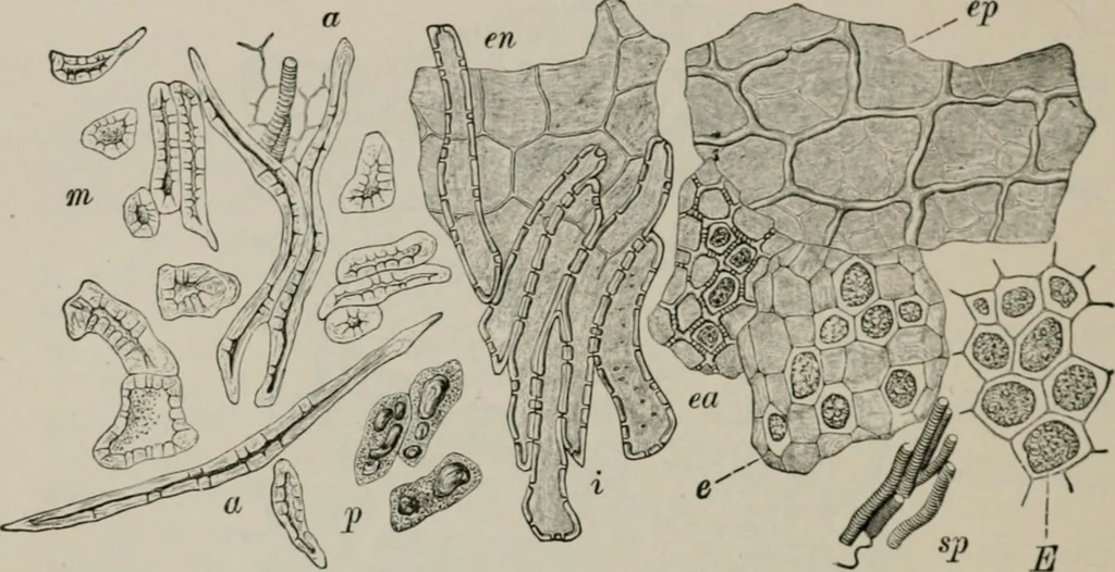
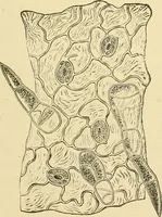
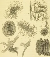
narrow lumen; occasional cells, however, have lumensbroader than the walls. An innermost layer (en) composed of compressed thin-walled paren-chyma cells lines the cavity. Spermoderm (Fig. 1S4). i. The Epidermal Cells (ep) seen in sur-face view are highly characteristic, owing to their unequally… … Fig. 184. Olive. I-Ilcmenls in surface view. /> oil cells of mesocarp; a, m, i stone cellsand fibers of endocarp; eu inner layer of endocarp; ep outer eindermis of sj)ermoderni;ea outer layer of endosperm; E and e parenchyma of cotyledon; sp spiral ves.*^*!. X i6o. (MOELLER.) and colorless…