The microscopy of vegetable foods, with special reference to the detection of adulteration and the diagnosis of mixtures (1916)
Winton, Andrew Lincoln, 1864-1946, Moeller, Josef, 1848-1924, Winton, Kate Grace Barber, 1882- · New York, John Wiley & sons, inc.; [etc., etc.] · leaf 265
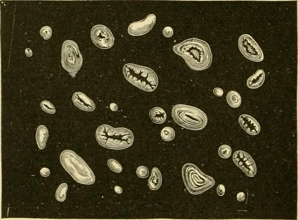
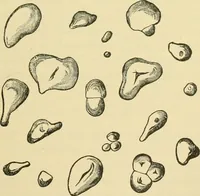
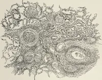
ass cells (subepidermallayer) s.nd p spongy parenchyma;c cotyledon with ep epidermis andaw starch cells. Xi6o. (Winton.) 3. Spongy Parenchyma (p). Fig. 196. Lentil. Hour-glasscells (subepidermal layer)of spermoderm in surface view. X 160. (MOELLER.) The Column Cells (sub) of hour-form are 18-35 P-… … Fig. 107. Lentil Starch. X300. (ISIoelllr.) cells are large, some of them containing a brown substance shov/ing thereaction for tannins. CHINA BEAN. 247 Embryo (Fig. 195, C). The thin-walled cells contain starch grains(Fig. 197) somewhat smaller than those in the bean or pea, the largestbeing but…
Plant anatomy · Food adulteration and inspection · Microscopy
Internet Archive · Library of Congress · Public Domain Mark 1.0. Free to use for anything.