The microscopy of vegetable foods, with special reference to the detection of adulteration and the diagnosis of mixtures (1916)
Winton, Andrew Lincoln, 1864-1946, Moeller, Josef, 1848-1924, Winton, Kate Grace Barber, 1882- · New York, John Wiley & sons, inc.; [etc., etc.] · leaf 279
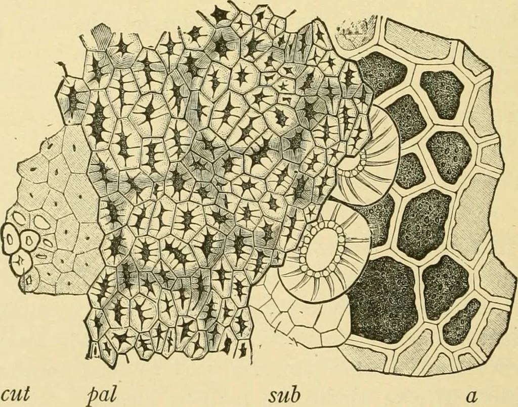
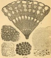
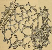
Fig. 213. Fenugreek {Trigonella Foenum-Graecum). FiG. 214. Fenugreek. Seed witha seed, natural size; the others enlarged. spermoderm partially removed (NOBBE.) showing cotyledon and radicle. (MOELLER.) The Spennoderm (Fig. 216, 5; Fig. 215) has the three layers of atypical legume. … Fig. 215. Fenugreek. Elements of seed in surface view, cut cuticle and blunt palisadecells; pal pointed pahsade cells; sub subepidermal cells and parenchyma; a aleuronecells of endosperm. (Tschirch.) I. The Palisade Cells (Figs, 215 and 216, pal), 60-75 A high and8-20 [1 broad, have narrow cavities…
Plant anatomy · Food adulteration and inspection · Microscopy
Internet Archive · Library of Congress · Public Domain Mark 1.0. Free to use for anything.