The microscopy of vegetable foods, with special reference to the detection of adulteration and the diagnosis of mixtures (1916)
Winton, Andrew Lincoln, 1864-1946, Moeller, Josef, 1848-1924, Winton, Kate Grace Barber, 1882- · New York, John Wiley & sons, inc.; [etc., etc.] · leaf 408
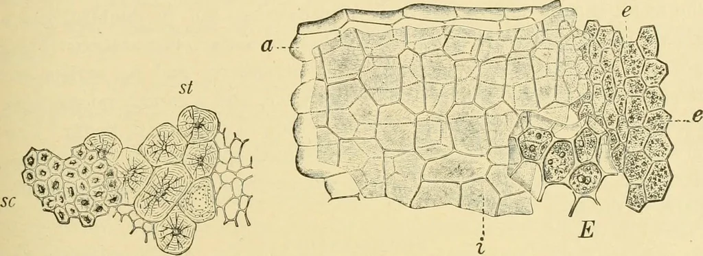
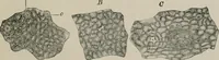
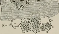
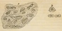
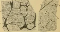
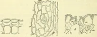
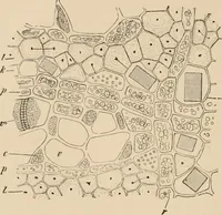
n diameter. They FIG. 389 have narrow lumen and thick walls with distinct rings and numerousbranching pores. They are readily distinguished from the smaller cellsof the outer sclerenchyma. Spermoderm (Fig. 302). The seed, which as a rule does not com-pletely fill the locule, is enveloped by a brown… … Fig, 301. Fig. Elements of peri-carp (shell of nutlet) in surfaceview, sc outer sclerenchyma layer;St stone cells of endocarp. X i6o. (MOELLER.) Fig. 302. Fig. Elements of seed in surface view.Spermoderm consists of a colorless outer epidermisand i brown inner layers; £ endosperm; e embryo. X160,…
Plant anatomy · Food adulteration and inspection · Microscopy
Internet Archive · Library of Congress · Public Domain Mark 1.0. Free to use for anything.