The microscopy of vegetable foods, with special reference to the detection of adulteration and the diagnosis of mixtures (1916)
Winton, Andrew Lincoln, 1864-1946, Moeller, Josef, 1848-1924, Winton, Kate Grace Barber, 1882- · New York, John Wiley & sons, inc.; [etc., etc.] · leaf 427
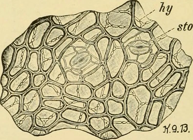
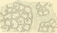
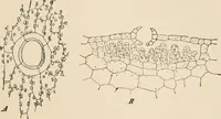
Tnes Fig. 312. Watermelon {CUridlus vulgaris). Pericarp in cross section, epi epicarp withsto stoma; hy hypoderm; st outer mesocarp (stone-cell layer); x parenchyma betweengroups of stone cells; we5 middle mesocarp. X160. (Barber.) … Fig. 312a. Watermelon. Pericarp in surface view. Epicarp with sto stoma; hy hypo-derm. X160. (Barber.) of looser texture, with numerous bundle fibers. Embedded in the innerpericarp are the black or light-brown, fiat, lustrous seeds. HISTOLOGY.Pericarp (Figs. 312, and 312a). i. The Epicarp {epi)…
Plant anatomy · Food adulteration and inspection · Microscopy
Internet Archive · Library of Congress · Public Domain Mark 1.0. Free to use for anything.