The microscopy of vegetable foods, with special reference to the detection of adulteration and the diagnosis of mixtures (1916)
Winton, Andrew Lincoln, 1864-1946, Moeller, Josef, 1848-1924, Winton, Kate Grace Barber, 1882- · New York, John Wiley & sons, inc.; [etc., etc.] · leaf 464
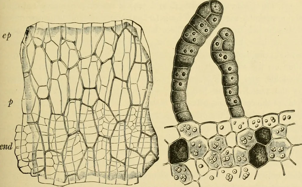
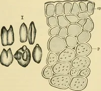
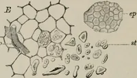
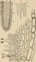
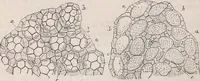
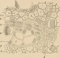
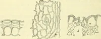
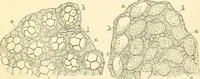
Numerous layers of spongy paren-chyma cells, through which pass the bundles of the raphe and its branches,form the third coat. Narrow spiral vessels readily separating fromthe other elements, characterize the bundles. 4. Slone Cells (Fig. 346, st; Fig. 349, d). The cells of the next layerare… … FlG. 347. Cocoa. Outer elements of shell. FiG. 348. Cocoa. Cross section of outer ep epidermis of spermoderm; end endocarp portion of cotyledon, showing hairs (cross cells); p parenchyma of mesocarp. (Mitscherlichian bodies) and starch X160, (MoELLER.) parenchyma. (Moeller.) encd at all,…
Plant anatomy · Food adulteration and inspection · Microscopy
Internet Archive · Library of Congress · Public Domain Mark 1.0. Free to use for anything.